Iron »
PDB 1gem-1gwe »
1gjn »
Iron in PDB 1gjn: Hydrogen Peroxide Derived Myoglobin Compound II at pH 5.2
Protein crystallography data
The structure of Hydrogen Peroxide Derived Myoglobin Compound II at pH 5.2, PDB code: 1gjn
was solved by
H.-P.Hersleth,
B.Dalhus,
C.H.Gorbitz,
K.K.Andersson,
with X-Ray Crystallography technique. A brief refinement statistics is given in the table below:
| Resolution Low / High (Å) | 20.79 / 1.35 |
| Space group | P 1 21 1 |
| Cell size a, b, c (Å), α, β, γ (°) | 62.862, 28.650, 35.352, 90.00, 105.93, 90.00 |
| R / Rfree (%) | 17.8 / 20.5 |
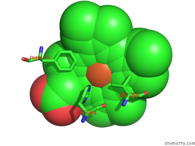
Iron Binding Sites:
The binding sites of Iron atom in the Hydrogen Peroxide Derived Myoglobin Compound II at pH 5.2
(pdb code 1gjn). This binding sites where shown within
5.0 Angstroms radius around Iron atom.
In total only one binding site of Iron was determined in the Hydrogen Peroxide Derived Myoglobin Compound II at pH 5.2, PDB code: 1gjn:
In total only one binding site of Iron was determined in the Hydrogen Peroxide Derived Myoglobin Compound II at pH 5.2, PDB code: 1gjn:
Iron binding site 1 out of 1 in 1gjn
Go back to
Iron binding site 1 out
of 1 in the Hydrogen Peroxide Derived Myoglobin Compound II at pH 5.2
Mono view

Stereo pair view
Mono view
Stereo pair view
A full contact list of Iron with other atoms in the Fe binding
site number 1 of Hydrogen Peroxide Derived Myoglobin Compound II at pH 5.2 within 5.0Å range:
|
Reference:
H.-P.Hersleth,
B.Dalhus,
C.H.Gorbitz,
K.K.Andersson.
An Iron Hydroxide Moiety in the 1.35 A Resolution Structure of Hydrogen Peroxide Derived Myoglobin Compound II at pH 5.2 J.Biol.Inorg.Chem. V. 7 299 2002.
ISSN: ISSN 0949-8257
PubMed: 11935353
DOI: 10.1007/S007750100296
Page generated: Wed Jul 16 14:59:10 2025
ISSN: ISSN 0949-8257
PubMed: 11935353
DOI: 10.1007/S007750100296
Last articles
Na in 8FSWNa in 8FSV
Na in 8FSU
Na in 8FST
Na in 8FSM
Na in 8FSN
Na in 8FSH
Na in 8FSG
Na in 8FSC
Na in 8FSF