Iron »
PDB 1gem-1gwe »
1gnt »
Iron in PDB 1gnt: Hybrid Cluster Protein From Desulfovibrio Vulgaris. X-Ray Structure at 1.25A Resolution Using Synchrotron Radiation.
Protein crystallography data
The structure of Hybrid Cluster Protein From Desulfovibrio Vulgaris. X-Ray Structure at 1.25A Resolution Using Synchrotron Radiation., PDB code: 1gnt
was solved by
S.Macedo,
E.P.Mitchell,
C.V.Romao,
S.J.Cooper,
R.Coelho,
M.Y.Liu,
A.V.Xavier,
J.Legall,
S.Bailey,
D.C.Garner,
W.R.Hagen,
M.Teixeira,
M.A.Carrondo,
P.Lindley,
with X-Ray Crystallography technique. A brief refinement statistics is given in the table below:
| Resolution Low / High (Å) | 19.92 / 1.25 |
| Space group | P 21 21 21 |
| Cell size a, b, c (Å), α, β, γ (°) | 63.810, 64.530, 151.870, 90.00, 90.00, 90.00 |
| R / Rfree (%) | 15.7 / 17.3 |
Iron Binding Sites:
The binding sites of Iron atom in the Hybrid Cluster Protein From Desulfovibrio Vulgaris. X-Ray Structure at 1.25A Resolution Using Synchrotron Radiation.
(pdb code 1gnt). This binding sites where shown within
5.0 Angstroms radius around Iron atom.
In total 8 binding sites of Iron where determined in the Hybrid Cluster Protein From Desulfovibrio Vulgaris. X-Ray Structure at 1.25A Resolution Using Synchrotron Radiation., PDB code: 1gnt:
Jump to Iron binding site number: 1; 2; 3; 4; 5; 6; 7; 8;
In total 8 binding sites of Iron where determined in the Hybrid Cluster Protein From Desulfovibrio Vulgaris. X-Ray Structure at 1.25A Resolution Using Synchrotron Radiation., PDB code: 1gnt:
Jump to Iron binding site number: 1; 2; 3; 4; 5; 6; 7; 8;
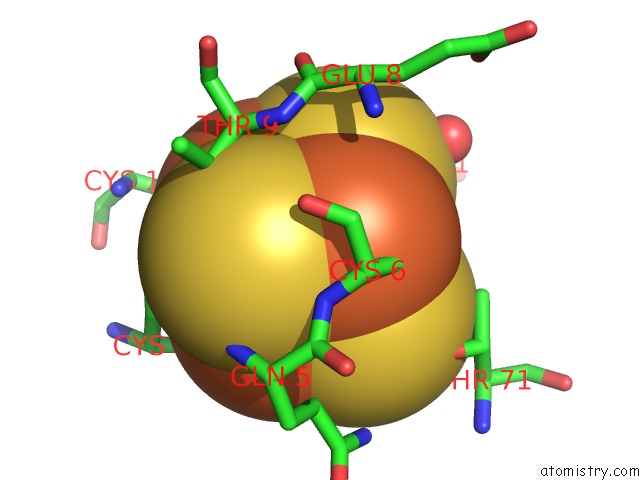
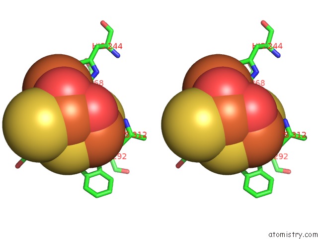
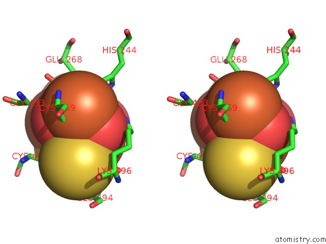
Iron binding site 1 out of 8 in 1gnt
Go back to
Iron binding site 1 out
of 8 in the Hybrid Cluster Protein From Desulfovibrio Vulgaris. X-Ray Structure at 1.25A Resolution Using Synchrotron Radiation.
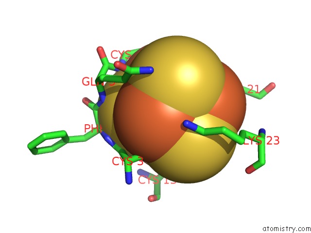
Mono view
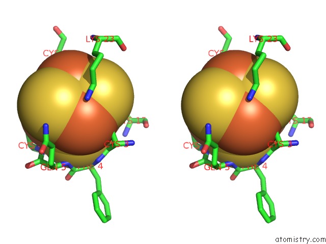
Stereo pair view
Mono view
Stereo pair view
A full contact list of Iron with other atoms in the Fe binding
site number 1 of Hybrid Cluster Protein From Desulfovibrio Vulgaris. X-Ray Structure at 1.25A Resolution Using Synchrotron Radiation. within 5.0Å range:
|
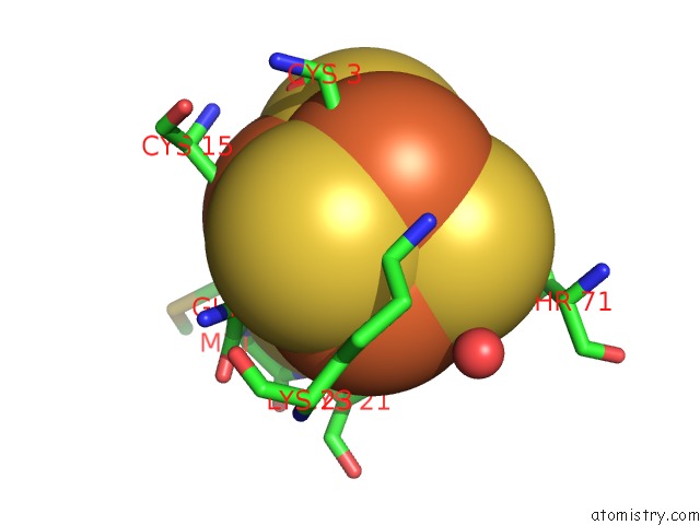
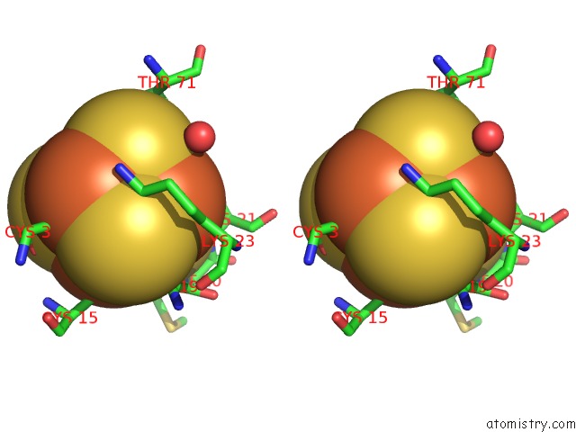
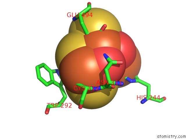
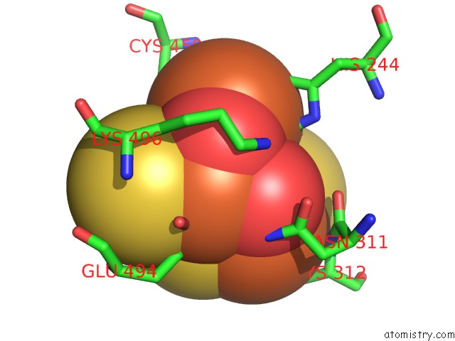
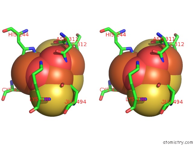
Iron binding site 2 out of 8 in 1gnt
Go back to
Iron binding site 2 out
of 8 in the Hybrid Cluster Protein From Desulfovibrio Vulgaris. X-Ray Structure at 1.25A Resolution Using Synchrotron Radiation.
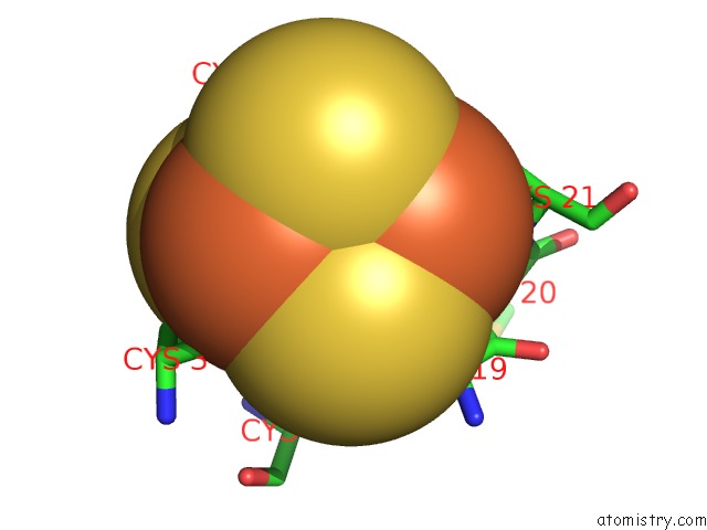
Mono view
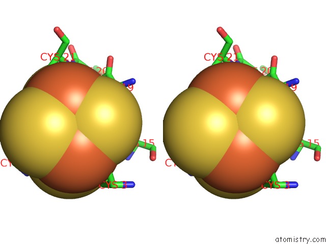
Stereo pair view
Mono view
Stereo pair view
A full contact list of Iron with other atoms in the Fe binding
site number 2 of Hybrid Cluster Protein From Desulfovibrio Vulgaris. X-Ray Structure at 1.25A Resolution Using Synchrotron Radiation. within 5.0Å range:
|
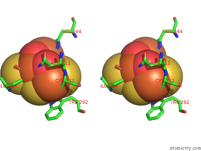
Iron binding site 3 out of 8 in 1gnt
Go back to
Iron binding site 3 out
of 8 in the Hybrid Cluster Protein From Desulfovibrio Vulgaris. X-Ray Structure at 1.25A Resolution Using Synchrotron Radiation.
Mono view

Stereo pair view
Mono view
Stereo pair view
A full contact list of Iron with other atoms in the Fe binding
site number 3 of Hybrid Cluster Protein From Desulfovibrio Vulgaris. X-Ray Structure at 1.25A Resolution Using Synchrotron Radiation. within 5.0Å range:
|
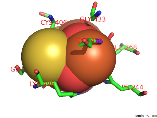
Iron binding site 4 out of 8 in 1gnt
Go back to
Iron binding site 4 out
of 8 in the Hybrid Cluster Protein From Desulfovibrio Vulgaris. X-Ray Structure at 1.25A Resolution Using Synchrotron Radiation.
Mono view
Stereo pair view
Mono view
Stereo pair view
A full contact list of Iron with other atoms in the Fe binding
site number 4 of Hybrid Cluster Protein From Desulfovibrio Vulgaris. X-Ray Structure at 1.25A Resolution Using Synchrotron Radiation. within 5.0Å range:
|
Iron binding site 5 out of 8 in 1gnt
Go back to
Iron binding site 5 out
of 8 in the Hybrid Cluster Protein From Desulfovibrio Vulgaris. X-Ray Structure at 1.25A Resolution Using Synchrotron Radiation.
Mono view
Stereo pair view
Mono view
Stereo pair view
A full contact list of Iron with other atoms in the Fe binding
site number 5 of Hybrid Cluster Protein From Desulfovibrio Vulgaris. X-Ray Structure at 1.25A Resolution Using Synchrotron Radiation. within 5.0Å range:
|
Iron binding site 6 out of 8 in 1gnt
Go back to
Iron binding site 6 out
of 8 in the Hybrid Cluster Protein From Desulfovibrio Vulgaris. X-Ray Structure at 1.25A Resolution Using Synchrotron Radiation.
Mono view
Stereo pair view
Mono view
Stereo pair view
A full contact list of Iron with other atoms in the Fe binding
site number 6 of Hybrid Cluster Protein From Desulfovibrio Vulgaris. X-Ray Structure at 1.25A Resolution Using Synchrotron Radiation. within 5.0Å range:
|
Iron binding site 7 out of 8 in 1gnt
Go back to
Iron binding site 7 out
of 8 in the Hybrid Cluster Protein From Desulfovibrio Vulgaris. X-Ray Structure at 1.25A Resolution Using Synchrotron Radiation.
Mono view
Stereo pair view
Mono view
Stereo pair view
A full contact list of Iron with other atoms in the Fe binding
site number 7 of Hybrid Cluster Protein From Desulfovibrio Vulgaris. X-Ray Structure at 1.25A Resolution Using Synchrotron Radiation. within 5.0Å range:
|
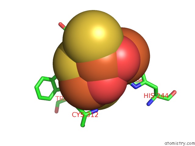
Iron binding site 8 out of 8 in 1gnt
Go back to
Iron binding site 8 out
of 8 in the Hybrid Cluster Protein From Desulfovibrio Vulgaris. X-Ray Structure at 1.25A Resolution Using Synchrotron Radiation.
Mono view
Stereo pair view
Mono view
Stereo pair view
A full contact list of Iron with other atoms in the Fe binding
site number 8 of Hybrid Cluster Protein From Desulfovibrio Vulgaris. X-Ray Structure at 1.25A Resolution Using Synchrotron Radiation. within 5.0Å range:
|
Reference:
S.Macedo,
E.P.Mitchell,
C.V.Romao,
S.J.Cooper,
R.Coelho,
M.Y.Liu,
A.V.Xavier,
J.Legall,
S.Bailey,
D.C.Garner,
W.R.Hagen,
M.Teixeira,
M.A.Carrondo,
P.Lindley.
Hybrid Cluster Proteins (Hcps) From Desulfovibrio Desulfuricans Atcc 27774 and Desulfovibrio Vulgaris (Hildenborough): X-Ray Structures at 1.25 A Resolution Using Synchrotron Radiation. J. Biol. Inorg. Chem. V. 7 514 2002.
ISSN: ISSN 0949-8257
PubMed: 11941509
DOI: 10.1007/S00775-001-0326-Y
Page generated: Wed Jul 16 15:02:29 2025
ISSN: ISSN 0949-8257
PubMed: 11941509
DOI: 10.1007/S00775-001-0326-Y
Last articles
Na in 8FPNNa in 8FPM
Na in 8FPD
Na in 8FPB
Na in 8FP8
Na in 8FP7
Na in 8FP6
Na in 8FHR
Na in 8FHT
Na in 8FF9