Iron »
PDB 1gwf-1h5f »
1gyo »
Iron in PDB 1gyo: Crystal Structure of the Di-Tetraheme Cytochrome C3 From Desulfovibrio Gigas at 1.2 Angstrom Resolution
Protein crystallography data
The structure of Crystal Structure of the Di-Tetraheme Cytochrome C3 From Desulfovibrio Gigas at 1.2 Angstrom Resolution, PDB code: 1gyo
was solved by
D.Aragao,
C.Frazao,
L.Sieker,
G.M.Sheldrick,
J.Legall,
M.A.Carrondo,
with X-Ray Crystallography technique. A brief refinement statistics is given in the table below:
| Resolution Low / High (Å) | 26.50 / 1.20 |
| Space group | P 31 |
| Cell size a, b, c (Å), α, β, γ (°) | 56.670, 56.670, 94.170, 90.00, 90.00, 120.00 |
| R / Rfree (%) | 13 / 15.7 |
Iron Binding Sites:
The binding sites of Iron atom in the Crystal Structure of the Di-Tetraheme Cytochrome C3 From Desulfovibrio Gigas at 1.2 Angstrom Resolution
(pdb code 1gyo). This binding sites where shown within
5.0 Angstroms radius around Iron atom.
In total 8 binding sites of Iron where determined in the Crystal Structure of the Di-Tetraheme Cytochrome C3 From Desulfovibrio Gigas at 1.2 Angstrom Resolution, PDB code: 1gyo:
Jump to Iron binding site number: 1; 2; 3; 4; 5; 6; 7; 8;
In total 8 binding sites of Iron where determined in the Crystal Structure of the Di-Tetraheme Cytochrome C3 From Desulfovibrio Gigas at 1.2 Angstrom Resolution, PDB code: 1gyo:
Jump to Iron binding site number: 1; 2; 3; 4; 5; 6; 7; 8;
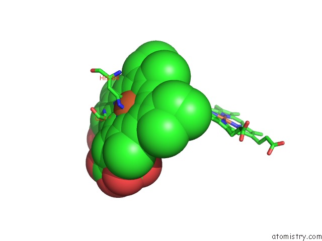
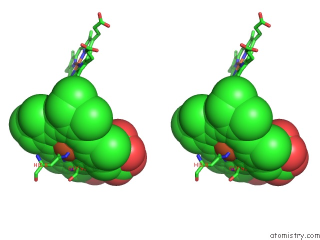
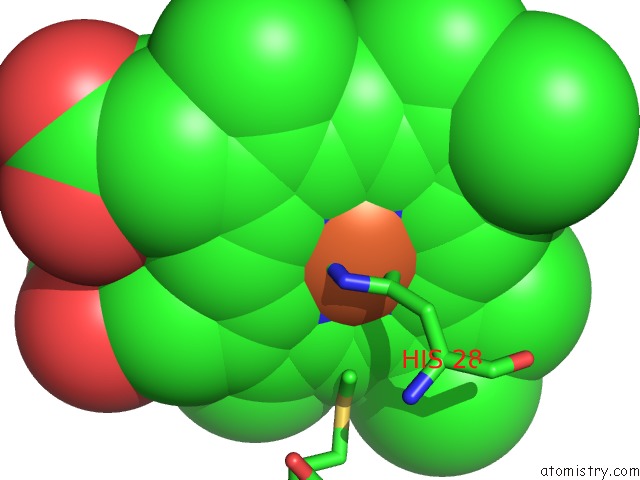
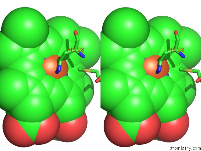
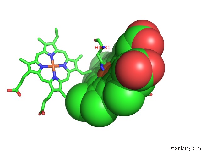
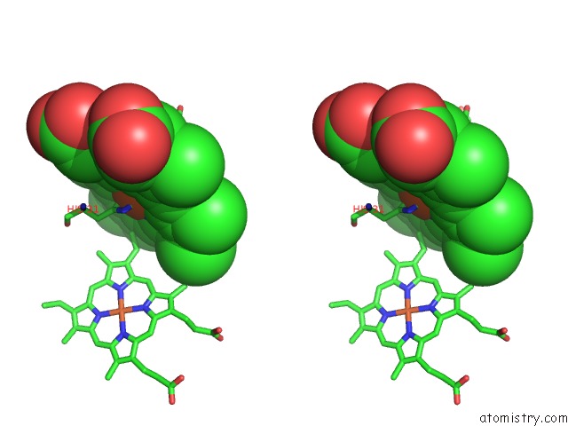
Iron binding site 1 out of 8 in 1gyo
Go back to
Iron binding site 1 out
of 8 in the Crystal Structure of the Di-Tetraheme Cytochrome C3 From Desulfovibrio Gigas at 1.2 Angstrom Resolution
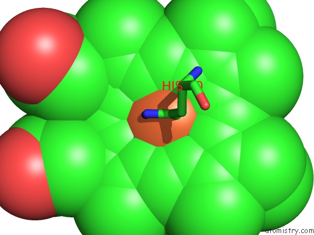
Mono view
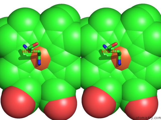
Stereo pair view
Mono view
Stereo pair view
A full contact list of Iron with other atoms in the Fe binding
site number 1 of Crystal Structure of the Di-Tetraheme Cytochrome C3 From Desulfovibrio Gigas at 1.2 Angstrom Resolution within 5.0Å range:
|
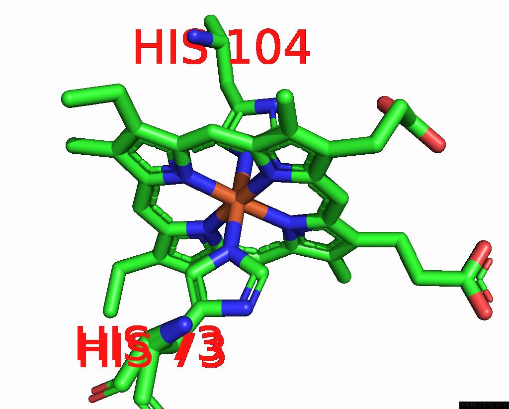
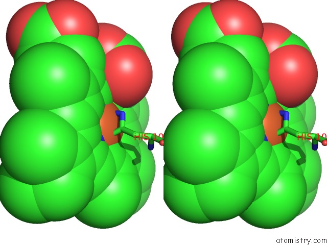
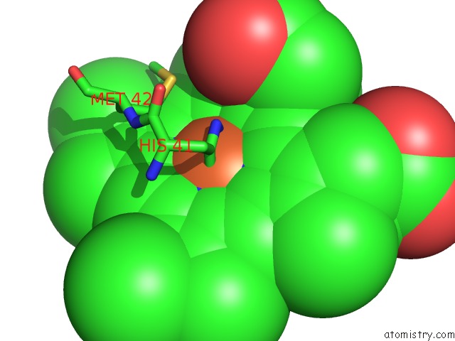
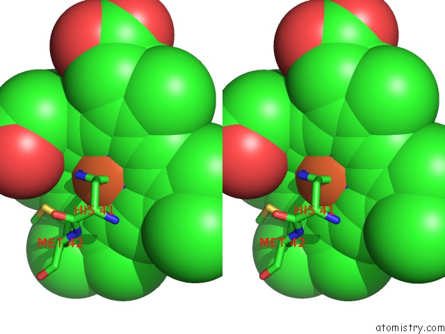
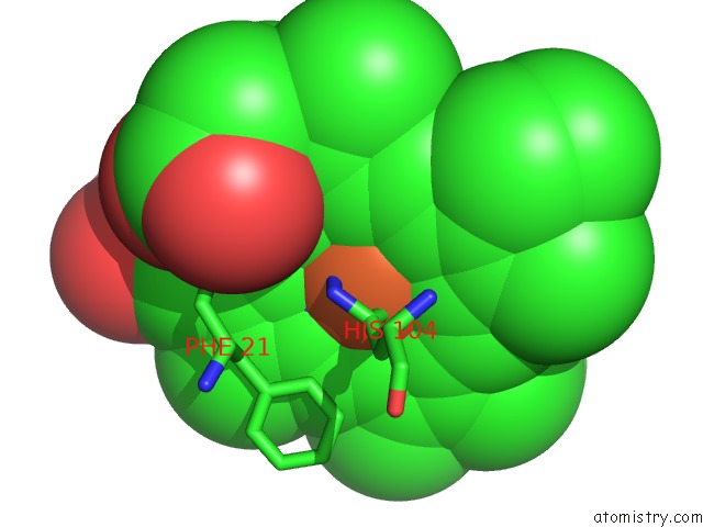
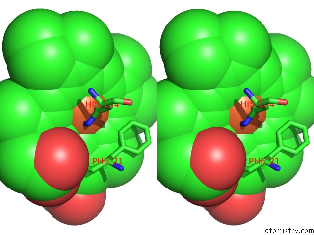
Iron binding site 2 out of 8 in 1gyo
Go back to
Iron binding site 2 out
of 8 in the Crystal Structure of the Di-Tetraheme Cytochrome C3 From Desulfovibrio Gigas at 1.2 Angstrom Resolution
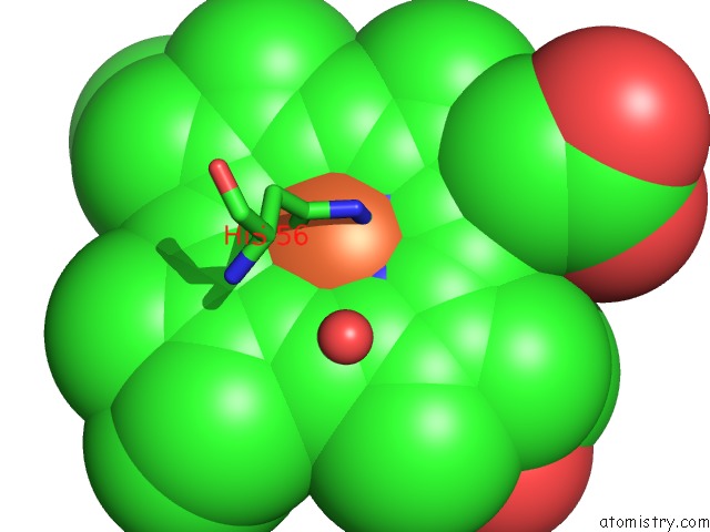
Mono view
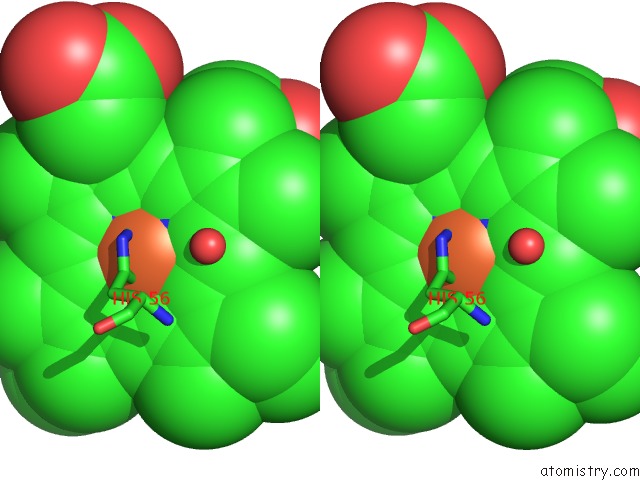
Stereo pair view
Mono view
Stereo pair view
A full contact list of Iron with other atoms in the Fe binding
site number 2 of Crystal Structure of the Di-Tetraheme Cytochrome C3 From Desulfovibrio Gigas at 1.2 Angstrom Resolution within 5.0Å range:
|
Iron binding site 3 out of 8 in 1gyo
Go back to
Iron binding site 3 out
of 8 in the Crystal Structure of the Di-Tetraheme Cytochrome C3 From Desulfovibrio Gigas at 1.2 Angstrom Resolution
Mono view
Stereo pair view
Mono view
Stereo pair view
A full contact list of Iron with other atoms in the Fe binding
site number 3 of Crystal Structure of the Di-Tetraheme Cytochrome C3 From Desulfovibrio Gigas at 1.2 Angstrom Resolution within 5.0Å range:
|
Iron binding site 4 out of 8 in 1gyo
Go back to
Iron binding site 4 out
of 8 in the Crystal Structure of the Di-Tetraheme Cytochrome C3 From Desulfovibrio Gigas at 1.2 Angstrom Resolution
Mono view
Stereo pair view
Mono view
Stereo pair view
A full contact list of Iron with other atoms in the Fe binding
site number 4 of Crystal Structure of the Di-Tetraheme Cytochrome C3 From Desulfovibrio Gigas at 1.2 Angstrom Resolution within 5.0Å range:
|
Iron binding site 5 out of 8 in 1gyo
Go back to
Iron binding site 5 out
of 8 in the Crystal Structure of the Di-Tetraheme Cytochrome C3 From Desulfovibrio Gigas at 1.2 Angstrom Resolution
Mono view
Stereo pair view
Mono view
Stereo pair view
A full contact list of Iron with other atoms in the Fe binding
site number 5 of Crystal Structure of the Di-Tetraheme Cytochrome C3 From Desulfovibrio Gigas at 1.2 Angstrom Resolution within 5.0Å range:
|
Iron binding site 6 out of 8 in 1gyo
Go back to
Iron binding site 6 out
of 8 in the Crystal Structure of the Di-Tetraheme Cytochrome C3 From Desulfovibrio Gigas at 1.2 Angstrom Resolution
Mono view
Stereo pair view
Mono view
Stereo pair view
A full contact list of Iron with other atoms in the Fe binding
site number 6 of Crystal Structure of the Di-Tetraheme Cytochrome C3 From Desulfovibrio Gigas at 1.2 Angstrom Resolution within 5.0Å range:
|
Iron binding site 7 out of 8 in 1gyo
Go back to
Iron binding site 7 out
of 8 in the Crystal Structure of the Di-Tetraheme Cytochrome C3 From Desulfovibrio Gigas at 1.2 Angstrom Resolution
Mono view
Stereo pair view
Mono view
Stereo pair view
A full contact list of Iron with other atoms in the Fe binding
site number 7 of Crystal Structure of the Di-Tetraheme Cytochrome C3 From Desulfovibrio Gigas at 1.2 Angstrom Resolution within 5.0Å range:
|
Iron binding site 8 out of 8 in 1gyo
Go back to
Iron binding site 8 out
of 8 in the Crystal Structure of the Di-Tetraheme Cytochrome C3 From Desulfovibrio Gigas at 1.2 Angstrom Resolution
Mono view
Stereo pair view
Mono view
Stereo pair view
A full contact list of Iron with other atoms in the Fe binding
site number 8 of Crystal Structure of the Di-Tetraheme Cytochrome C3 From Desulfovibrio Gigas at 1.2 Angstrom Resolution within 5.0Å range:
|
Reference:
D.Aragao,
C.Frazao,
L.Sieker,
G.M.Sheldrick,
J.Legall,
M.A.Carrondo.
Structure of Dimeric Cytochrome C3 From Desulfovibrio Gigas at 1.2 A Resolution Acta Crystallogr.,Sect.D V. 59 644 2003.
ISSN: ISSN 0907-4449
PubMed: 12657783
DOI: 10.1107/S090744490300194X
Page generated: Wed Jul 16 15:22:59 2025
ISSN: ISSN 0907-4449
PubMed: 12657783
DOI: 10.1107/S090744490300194X
Last articles
Fe in 6ZKLFe in 6ZKI
Fe in 6ZKK
Fe in 6ZKJ
Fe in 6ZKH
Fe in 6ZKG
Fe in 6ZKF
Fe in 6ZKE
Fe in 6ZKD
Fe in 6ZKC