Atomistry » Iron » PDB 1h5g-1hdb » 1hbs
Atomistry »
  Iron »
    PDB 1h5g-1hdb »
      1hbs »

Iron in PDB 1hbs: Refined Crystal Structure of Deoxyhemoglobin S. I. Restrained Least-Squares Refinement at 3.0-Angstroms Resolution

Protein crystallography data

The structure of Refined Crystal Structure of Deoxyhemoglobin S. I. Restrained Least-Squares Refinement at 3.0-Angstroms Resolution, PDB code: 1hbs was solved by E.A.Padlan, W.E.Love, with X-Ray Crystallography technique. A brief refinement statistics is given in the table below:

Resolution Low / High (Å) N/A / 3.00
Space group P 1 21 1
Cell size a, b, c (Å), α, β, γ (°) 63.330, 185.660, 52.970, 90.00, 92.69, 90.00
R / Rfree (%) 25.4 / n/a

Iron Binding Sites:

The binding sites of Iron atom in the Refined Crystal Structure of Deoxyhemoglobin S. I. Restrained Least-Squares Refinement at 3.0-Angstroms Resolution (pdb code 1hbs). This binding sites where shown within 5.0 Angstroms radius around Iron atom.
In total 8 binding sites of Iron where determined in the Refined Crystal Structure of Deoxyhemoglobin S. I. Restrained Least-Squares Refinement at 3.0-Angstroms Resolution, PDB code: 1hbs:
Jump to Iron binding site number: 1; 2; 3; 4; 5; 6; 7; 8;

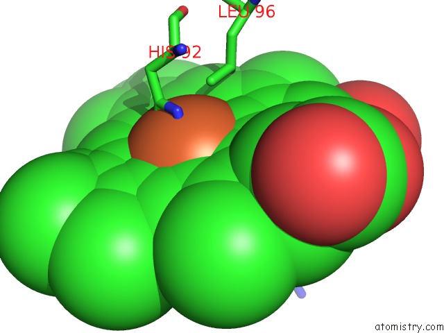
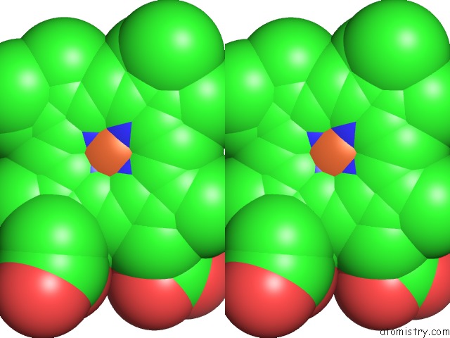
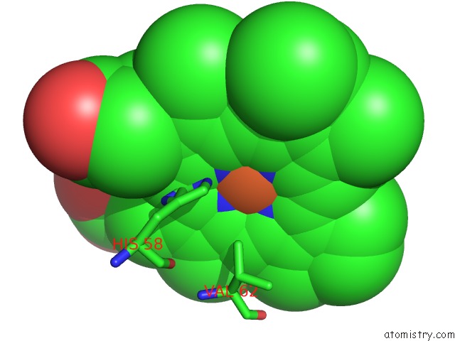
Iron binding site 1 out of 8 in 1hbs

Go back to Iron Binding Sites List in 1hbs
Iron binding site 1 out of 8 in the Refined Crystal Structure of Deoxyhemoglobin S. I. Restrained Least-Squares Refinement at 3.0-Angstroms Resolution


Mono view


Stereo pair view

A full contact list of Iron with other atoms in the Fe binding site number 1 of Refined Crystal Structure of Deoxyhemoglobin S. I. Restrained Least-Squares Refinement at 3.0-Angstroms Resolution within 5.0Å range:
probe atom residue distance (Å) B Occ
A:Fe142

b:23.5
occ:1.00
FE A:HEM142 0.0 23.5 1.0
NC A:HEM142 2.0 22.1 1.0
NB A:HEM142 2.0 20.6 1.0
ND A:HEM142 2.0 22.9 1.0
NA A:HEM142 2.2 21.6 1.0
NE2 A:HIS87 2.4 26.0 1.0
C4D A:HEM142 3.0 21.1 1.0
C4B A:HEM142 3.0 20.3 1.0
C1B A:HEM142 3.0 21.0 1.0
C4C A:HEM142 3.0 21.9 1.0
C1A A:HEM142 3.1 21.4 1.0
C4A A:HEM142 3.2 20.6 1.0
C1D A:HEM142 3.2 21.7 1.0
CE1 A:HIS87 3.2 27.7 1.0
C1C A:HEM142 3.3 21.8 1.0
CHD A:HEM142 3.3 22.7 1.0
CD2 A:HIS87 3.4 24.6 1.0
CHB A:HEM142 3.4 21.0 1.0
CHA A:HEM142 3.5 22.1 1.0
CHC A:HEM142 3.6 22.3 1.0
C2B A:HEM142 4.2 20.2 1.0
C3B A:HEM142 4.2 19.4 1.0
C3D A:HEM142 4.2 19.6 1.0
C2C A:HEM142 4.4 20.7 1.0
C3C A:HEM142 4.4 20.4 1.0
C2D A:HEM142 4.4 20.1 1.0
C2A A:HEM142 4.4 20.7 1.0
ND1 A:HIS87 4.5 25.9 1.0
CG A:HIS87 4.5 24.1 1.0
C3A A:HEM142 4.5 20.7 1.0
CZ A:PHE98 4.8 23.1 1.0

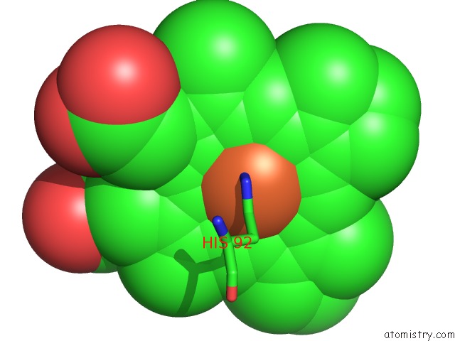
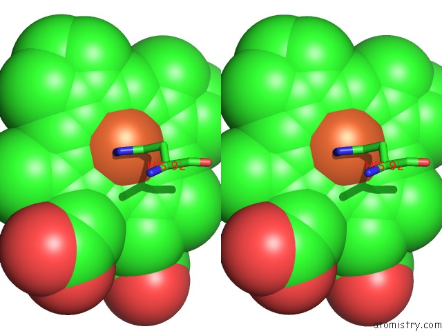
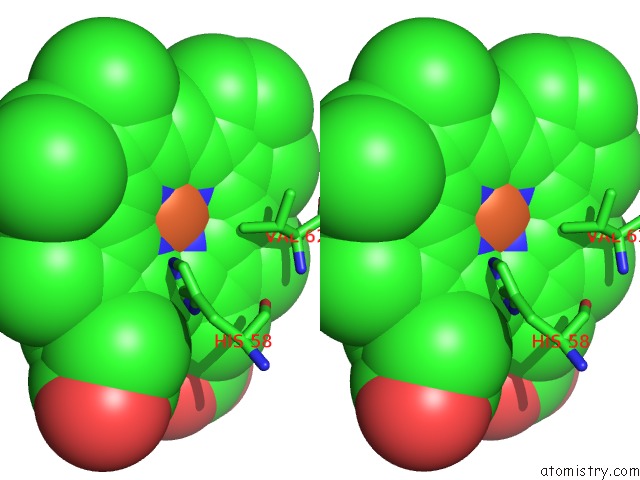
Iron binding site 2 out of 8 in 1hbs

Go back to Iron Binding Sites List in 1hbs
Iron binding site 2 out of 8 in the Refined Crystal Structure of Deoxyhemoglobin S. I. Restrained Least-Squares Refinement at 3.0-Angstroms Resolution


Mono view


Stereo pair view

A full contact list of Iron with other atoms in the Fe binding site number 2 of Refined Crystal Structure of Deoxyhemoglobin S. I. Restrained Least-Squares Refinement at 3.0-Angstroms Resolution within 5.0Å range:
probe atom residue distance (Å) B Occ
B:Fe147

b:21.8
occ:1.00
FE B:HEM147 0.0 21.8 1.0
NA B:HEM147 1.9 19.4 1.0
NE2 B:HIS92 2.0 16.0 1.0
NC B:HEM147 2.0 20.1 1.0
NB B:HEM147 2.1 17.4 1.0
ND B:HEM147 2.2 20.8 1.0
CE1 B:HIS92 2.7 14.6 1.0
CD2 B:HIS92 2.8 14.9 1.0
C4A B:HEM147 2.9 19.7 1.0
C4D B:HEM147 2.9 20.1 1.0
C1A B:HEM147 3.0 18.3 1.0
C1B B:HEM147 3.0 18.6 1.0
C4C B:HEM147 3.1 20.3 1.0
C1C B:HEM147 3.1 20.6 1.0
C4B B:HEM147 3.2 17.7 1.0
CHA B:HEM147 3.4 18.3 1.0
CHB B:HEM147 3.4 20.2 1.0
CHC B:HEM147 3.5 20.7 1.0
C1D B:HEM147 3.5 18.6 1.0
ND1 B:HIS92 3.7 14.3 1.0
CG B:HIS92 3.9 15.2 1.0
CHD B:HEM147 3.9 19.7 1.0
C2A B:HEM147 4.0 19.2 1.0
C3A B:HEM147 4.0 19.9 1.0
C2C B:HEM147 4.2 18.4 1.0
C3D B:HEM147 4.2 20.3 1.0
C2B B:HEM147 4.2 17.6 1.0
C3C B:HEM147 4.3 18.2 1.0
C3B B:HEM147 4.3 16.9 1.0
CD1 B:LEU96 4.3 24.7 1.0
C2D B:HEM147 4.5 19.9 1.0
NE2 B:HIS63 4.7 24.7 1.0
CE1 B:HIS63 4.9 24.0 1.0

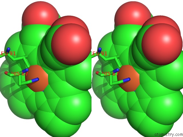
Iron binding site 3 out of 8 in 1hbs

Go back to Iron Binding Sites List in 1hbs
Iron binding site 3 out of 8 in the Refined Crystal Structure of Deoxyhemoglobin S. I. Restrained Least-Squares Refinement at 3.0-Angstroms Resolution


Mono view


Stereo pair view

A full contact list of Iron with other atoms in the Fe binding site number 3 of Refined Crystal Structure of Deoxyhemoglobin S. I. Restrained Least-Squares Refinement at 3.0-Angstroms Resolution within 5.0Å range:
probe atom residue distance (Å) B Occ
C:Fe142

b:17.3
occ:1.00
FE C:HEM142 0.0 17.3 1.0
NC C:HEM142 2.1 12.5 1.0
NA C:HEM142 2.1 16.6 1.0
NB C:HEM142 2.1 12.6 1.0
ND C:HEM142 2.1 15.1 1.0
NE2 C:HIS87 2.5 8.6 1.0
CD2 C:HIS87 2.9 10.8 1.0
C1B C:HEM142 3.0 12.4 1.0
C1A C:HEM142 3.0 18.3 1.0
C1D C:HEM142 3.1 16.4 1.0
C4C C:HEM142 3.1 13.5 1.0
C4D C:HEM142 3.1 17.3 1.0
C1C C:HEM142 3.1 11.3 1.0
C4B C:HEM142 3.1 11.4 1.0
C4A C:HEM142 3.1 16.8 1.0
CHB C:HEM142 3.3 14.5 1.0
CHA C:HEM142 3.5 18.0 1.0
CHC C:HEM142 3.5 11.0 1.0
CHD C:HEM142 3.5 15.6 1.0
CE1 C:HIS87 3.7 8.1 1.0
CG C:HIS87 4.2 11.3 1.0
C3B C:HEM142 4.3 11.1 1.0
C3C C:HEM142 4.3 12.0 1.0
C2B C:HEM142 4.3 12.1 1.0
C2C C:HEM142 4.3 10.1 1.0
C2A C:HEM142 4.3 19.3 1.0
C3D C:HEM142 4.3 18.3 1.0
C3A C:HEM142 4.3 18.0 1.0
C2D C:HEM142 4.3 18.5 1.0
ND1 C:HIS87 4.5 9.7 1.0

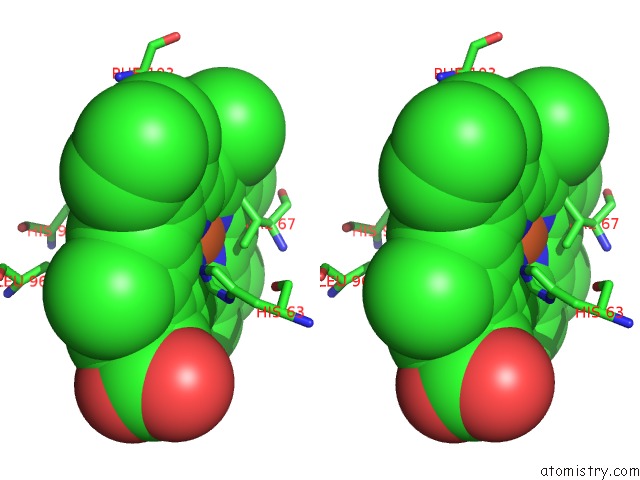
Iron binding site 4 out of 8 in 1hbs

Go back to Iron Binding Sites List in 1hbs
Iron binding site 4 out of 8 in the Refined Crystal Structure of Deoxyhemoglobin S. I. Restrained Least-Squares Refinement at 3.0-Angstroms Resolution


Mono view


Stereo pair view

A full contact list of Iron with other atoms in the Fe binding site number 4 of Refined Crystal Structure of Deoxyhemoglobin S. I. Restrained Least-Squares Refinement at 3.0-Angstroms Resolution within 5.0Å range:
probe atom residue distance (Å) B Occ
D:Fe147

b:35.2
occ:1.00
FE D:HEM147 0.0 35.2 1.0
NC D:HEM147 2.1 36.1 1.0
NE2 D:HIS92 2.1 26.8 1.0
NB D:HEM147 2.1 32.9 1.0
NA D:HEM147 2.1 33.9 1.0
ND D:HEM147 2.1 34.2 1.0
CE1 D:HIS92 2.9 26.7 1.0
C4D D:HEM147 3.0 33.1 1.0
C1B D:HEM147 3.0 33.2 1.0
C1D D:HEM147 3.1 35.5 1.0
C4B D:HEM147 3.1 32.2 1.0
C1A D:HEM147 3.1 33.1 1.0
CD2 D:HIS92 3.1 26.1 1.0
C1C D:HEM147 3.1 36.1 1.0
C4A D:HEM147 3.2 33.4 1.0
C4C D:HEM147 3.2 34.7 1.0
CHC D:HEM147 3.4 33.8 1.0
CHA D:HEM147 3.5 31.8 1.0
CHB D:HEM147 3.5 33.2 1.0
CHD D:HEM147 3.6 35.5 1.0
CG1 D:VAL67 3.6 8.7 1.0
NE2 D:HIS63 4.0 24.4 1.0
ND1 D:HIS92 4.1 27.3 1.0
CG D:HIS92 4.2 26.4 1.0
C3B D:HEM147 4.2 33.3 1.0
C2B D:HEM147 4.2 33.5 1.0
CE1 D:HIS63 4.3 24.4 1.0
C2D D:HEM147 4.3 35.3 1.0
C3D D:HEM147 4.3 34.8 1.0
C2A D:HEM147 4.3 33.2 1.0
C3C D:HEM147 4.3 34.2 1.0
C2C D:HEM147 4.4 35.9 1.0
C3A D:HEM147 4.4 33.4 1.0
CB D:VAL67 4.6 13.7 1.0
CG2 D:VAL67 4.9 11.0 1.0

Iron binding site 5 out of 8 in 1hbs

Go back to Iron Binding Sites List in 1hbs
Iron binding site 5 out of 8 in the Refined Crystal Structure of Deoxyhemoglobin S. I. Restrained Least-Squares Refinement at 3.0-Angstroms Resolution


Mono view


Stereo pair view

A full contact list of Iron with other atoms in the Fe binding site number 5 of Refined Crystal Structure of Deoxyhemoglobin S. I. Restrained Least-Squares Refinement at 3.0-Angstroms Resolution within 5.0Å range:
probe atom residue distance (Å) B Occ
E:Fe142

b:30.0
occ:1.00
FE E:HEM142 0.0 30.0 1.0
NC E:HEM142 1.9 26.9 1.0
NA E:HEM142 2.1 26.1 1.0
ND E:HEM142 2.1 24.9 1.0
NB E:HEM142 2.1 28.1 1.0
NE2 E:HIS87 2.6 21.6 1.0
C1C E:HEM142 2.9 24.0 1.0
C1D E:HEM142 2.9 24.6 1.0
C4C E:HEM142 3.0 25.4 1.0
C4A E:HEM142 3.0 26.9 1.0
C4B E:HEM142 3.0 26.5 1.0
CHD E:HEM142 3.1 24.6 1.0
C1B E:HEM142 3.1 28.5 1.0
CD2 E:HIS87 3.2 19.5 1.0
C1A E:HEM142 3.2 27.3 1.0
C4D E:HEM142 3.2 26.4 1.0
CHC E:HEM142 3.3 24.6 1.0
CHB E:HEM142 3.6 27.6 1.0
CHA E:HEM142 3.6 28.1 1.0
CE1 E:HIS87 3.7 23.0 1.0
C2C E:HEM142 4.0 22.6 1.0
C3C E:HEM142 4.1 23.9 1.0
NE2 E:HIS58 4.2 9.6 1.0
C3B E:HEM142 4.2 27.6 1.0
C2D E:HEM142 4.2 24.5 1.0
C2B E:HEM142 4.3 27.2 1.0
C3D E:HEM142 4.3 25.6 1.0
C3A E:HEM142 4.4 27.5 1.0
CG E:HIS87 4.4 20.1 1.0
C2A E:HEM142 4.5 27.0 1.0
ND1 E:HIS87 4.7 22.8 1.0
CE1 E:HIS58 4.8 6.8 1.0
CG2 E:VAL62 5.0 20.5 1.0

Iron binding site 6 out of 8 in 1hbs

Go back to Iron Binding Sites List in 1hbs
Iron binding site 6 out of 8 in the Refined Crystal Structure of Deoxyhemoglobin S. I. Restrained Least-Squares Refinement at 3.0-Angstroms Resolution


Mono view


Stereo pair view

A full contact list of Iron with other atoms in the Fe binding site number 6 of Refined Crystal Structure of Deoxyhemoglobin S. I. Restrained Least-Squares Refinement at 3.0-Angstroms Resolution within 5.0Å range:
probe atom residue distance (Å) B Occ
F:Fe147

b:25.9
occ:1.00
FE F:HEM147 0.0 25.9 1.0
NB F:HEM147 1.9 25.5 1.0
ND F:HEM147 2.0 26.7 1.0
NC F:HEM147 2.0 24.5 1.0
NA F:HEM147 2.2 29.3 1.0
NE2 F:HIS92 2.6 22.6 1.0
C4B F:HEM147 2.8 24.7 1.0
C4C F:HEM147 2.9 24.6 1.0
C4D F:HEM147 3.0 27.4 1.0
C1B F:HEM147 3.0 25.8 1.0
C1A F:HEM147 3.1 29.2 1.0
C1D F:HEM147 3.1 26.4 1.0
C1C F:HEM147 3.1 24.0 1.0
CE1 F:HIS92 3.2 22.8 1.0
CHA F:HEM147 3.4 28.3 1.0
CG2 F:VAL67 3.4 18.3 1.0
CHB F:HEM147 3.4 28.9 1.0
C4A F:HEM147 3.4 29.6 1.0
CHD F:HEM147 3.5 26.9 1.0
CHC F:HEM147 3.6 23.3 1.0
CD2 F:HIS92 3.7 21.8 1.0
CE1 F:HIS63 3.8 21.9 1.0
C3B F:HEM147 4.1 24.1 1.0
C2B F:HEM147 4.1 24.2 1.0
C3D F:HEM147 4.3 27.7 1.0
C2D F:HEM147 4.3 24.9 1.0
NE2 F:HIS63 4.3 22.0 1.0
C3C F:HEM147 4.3 21.8 1.0
C2C F:HEM147 4.3 22.3 1.0
ND1 F:HIS92 4.4 21.4 1.0
C2A F:HEM147 4.5 30.5 1.0
C3A F:HEM147 4.5 31.1 1.0
ND1 F:HIS63 4.6 23.3 1.0
CB F:VAL67 4.7 16.9 1.0
CG F:HIS92 4.8 21.1 1.0

Iron binding site 7 out of 8 in 1hbs

Go back to Iron Binding Sites List in 1hbs
Iron binding site 7 out of 8 in the Refined Crystal Structure of Deoxyhemoglobin S. I. Restrained Least-Squares Refinement at 3.0-Angstroms Resolution


Mono view


Stereo pair view

A full contact list of Iron with other atoms in the Fe binding site number 7 of Refined Crystal Structure of Deoxyhemoglobin S. I. Restrained Least-Squares Refinement at 3.0-Angstroms Resolution within 5.0Å range:
probe atom residue distance (Å) B Occ
G:Fe142

b:22.7
occ:1.00
FE G:HEM142 0.0 22.7 1.0
NC G:HEM142 1.9 24.9 1.0
NA G:HEM142 1.9 23.0 1.0
ND G:HEM142 2.1 24.1 1.0
NB G:HEM142 2.3 23.2 1.0
NE2 G:HIS87 2.7 4.7 1.0
CE1 G:HIS87 2.7 4.4 1.0
C1A G:HEM142 2.8 22.1 1.0
C4C G:HEM142 3.0 26.3 1.0
C1C G:HEM142 3.0 24.7 1.0
C4D G:HEM142 3.0 25.7 1.0
C1D G:HEM142 3.0 24.6 1.0
C4A G:HEM142 3.1 24.6 1.0
CHA G:HEM142 3.2 23.1 1.0
C4B G:HEM142 3.3 22.7 1.0
CHD G:HEM142 3.3 24.5 1.0
C1B G:HEM142 3.4 22.2 1.0
CHC G:HEM142 3.5 24.4 1.0
CE1 G:HIS58 3.6 42.1 1.0
CHB G:HEM142 3.8 22.8 1.0
CD2 G:HIS87 4.0 3.5 1.0
ND1 G:HIS87 4.0 4.4 1.0
C2A G:HEM142 4.1 23.6 1.0
C2C G:HEM142 4.2 25.2 1.0
C3C G:HEM142 4.2 27.0 1.0
C3A G:HEM142 4.3 24.2 1.0
C3D G:HEM142 4.3 27.8 1.0
C2D G:HEM142 4.4 26.2 1.0
ND1 G:HIS58 4.4 40.7 1.0
CD2 G:LEU91 4.4 19.0 1.0
NE2 G:HIS58 4.4 42.0 1.0
C3B G:HEM142 4.5 21.5 1.0
CD1 G:LEU91 4.5 17.8 1.0
C2B G:HEM142 4.6 22.3 1.0
CG G:HIS87 4.7 6.6 1.0

Iron binding site 8 out of 8 in 1hbs

Go back to Iron Binding Sites List in 1hbs
Iron binding site 8 out of 8 in the Refined Crystal Structure of Deoxyhemoglobin S. I. Restrained Least-Squares Refinement at 3.0-Angstroms Resolution


Mono view


Stereo pair view

A full contact list of Iron with other atoms in the Fe binding site number 8 of Refined Crystal Structure of Deoxyhemoglobin S. I. Restrained Least-Squares Refinement at 3.0-Angstroms Resolution within 5.0Å range:
probe atom residue distance (Å) B Occ
H:Fe147

b:26.8
occ:1.00
FE H:HEM147 0.0 26.8 1.0
NB H:HEM147 2.0 23.9 1.0
NA H:HEM147 2.1 25.6 1.0
NC H:HEM147 2.1 23.4 1.0
ND H:HEM147 2.1 24.3 1.0
CE1 H:HIS92 2.4 14.9 1.0
NE2 H:HIS92 2.8 17.8 1.0
C1C H:HEM147 3.0 21.6 1.0
C1D H:HEM147 3.0 24.6 1.0
C1B H:HEM147 3.0 23.3 1.0
C4A H:HEM147 3.0 24.5 1.0
C4B H:HEM147 3.1 22.5 1.0
C1A H:HEM147 3.1 25.8 1.0
C4D H:HEM147 3.2 24.4 1.0
C4C H:HEM147 3.2 23.7 1.0
CHC H:HEM147 3.2 20.9 1.0
CHB H:HEM147 3.4 23.5 1.0
CHD H:HEM147 3.5 24.8 1.0
CHA H:HEM147 3.5 24.1 1.0
ND1 H:HIS92 3.6 12.4 1.0
CD2 H:HIS92 4.1 15.5 1.0
C2C H:HEM147 4.1 22.7 1.0
C2B H:HEM147 4.2 22.5 1.0
C3B H:HEM147 4.2 22.8 1.0
C3A H:HEM147 4.2 24.9 1.0
C3C H:HEM147 4.3 22.9 1.0
C2A H:HEM147 4.3 26.3 1.0
C2D H:HEM147 4.3 22.9 1.0
NE2 H:HIS63 4.4 16.2 1.0
CG H:HIS92 4.4 14.4 1.0
C3D H:HEM147 4.4 23.3 1.0
CE1 H:PHE103 4.5 15.8 1.0
CG1 H:VAL67 4.7 20.4 1.0
CG2 H:VAL67 4.7 19.8 1.0
CE1 H:HIS63 4.7 14.3 1.0
CD1 H:LEU96 4.7 11.4 1.0

Reference:

E.A.Padlan, W.E.Love. Refined Crystal Structure of Deoxyhemoglobin S. I. Restrained Least-Squares Refinement at 3.0-A Resolution. J.Biol.Chem. V. 260 8272 1985.
ISSN: ISSN 0021-9258
PubMed: 4008491
Page generated: Wed Jul 16 15:51:06 2025

Last articles

F in 9VCL
F in 9VCK
F in 9JHS
F in 9JF9
F in 9CPK
Cu in 9OH6
Cu in 9OH7
Cu in 9VQM
Cl in 9QM5
Cl in 9RM2
© Copyright 2008-2020 by atomistry.com
Home   |    Site Map   |    Copyright   |    Contact us   |    Privacy