Iron »
PDB 1h5g-1hdb »
1hdb »
Iron in PDB 1hdb: Analysis of the Crystal Structure, Molecular Modeling and Infrared Spectroscopy of the Distal Beta-Heme Pocket VALINE67(E11)-Threonine Mutation of Hemoglobin
Protein crystallography data
The structure of Analysis of the Crystal Structure, Molecular Modeling and Infrared Spectroscopy of the Distal Beta-Heme Pocket VALINE67(E11)-Threonine Mutation of Hemoglobin, PDB code: 1hdb
was solved by
I.Pechik,
X.Ji,
C.Fronticelli,
G.L.Gilliland,
with X-Ray Crystallography technique. A brief refinement statistics is given in the table below:
| Resolution Low / High (Å) | 6.00 / 2.20 |
| Space group | P 1 21 1 |
| Cell size a, b, c (Å), α, β, γ (°) | 63.540, 83.190, 54.020, 90.00, 99.15, 90.00 |
| R / Rfree (%) | n/a / n/a |
Iron Binding Sites:
The binding sites of Iron atom in the Analysis of the Crystal Structure, Molecular Modeling and Infrared Spectroscopy of the Distal Beta-Heme Pocket VALINE67(E11)-Threonine Mutation of Hemoglobin
(pdb code 1hdb). This binding sites where shown within
5.0 Angstroms radius around Iron atom.
In total 4 binding sites of Iron where determined in the Analysis of the Crystal Structure, Molecular Modeling and Infrared Spectroscopy of the Distal Beta-Heme Pocket VALINE67(E11)-Threonine Mutation of Hemoglobin, PDB code: 1hdb:
Jump to Iron binding site number: 1; 2; 3; 4;
In total 4 binding sites of Iron where determined in the Analysis of the Crystal Structure, Molecular Modeling and Infrared Spectroscopy of the Distal Beta-Heme Pocket VALINE67(E11)-Threonine Mutation of Hemoglobin, PDB code: 1hdb:
Jump to Iron binding site number: 1; 2; 3; 4;
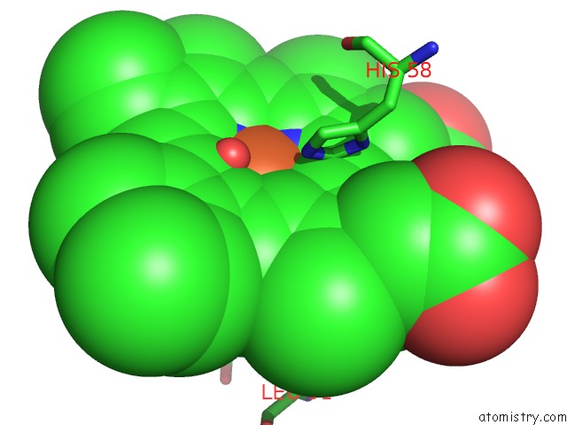
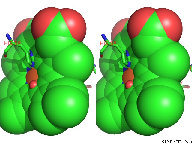
Iron binding site 1 out of 4 in 1hdb
Go back to
Iron binding site 1 out
of 4 in the Analysis of the Crystal Structure, Molecular Modeling and Infrared Spectroscopy of the Distal Beta-Heme Pocket VALINE67(E11)-Threonine Mutation of Hemoglobin
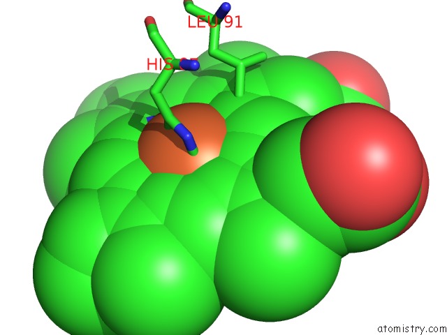
Mono view
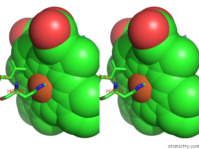
Stereo pair view
Mono view
Stereo pair view
A full contact list of Iron with other atoms in the Fe binding
site number 1 of Analysis of the Crystal Structure, Molecular Modeling and Infrared Spectroscopy of the Distal Beta-Heme Pocket VALINE67(E11)-Threonine Mutation of Hemoglobin within 5.0Å range:
|
Iron binding site 2 out of 4 in 1hdb
Go back to
Iron binding site 2 out
of 4 in the Analysis of the Crystal Structure, Molecular Modeling and Infrared Spectroscopy of the Distal Beta-Heme Pocket VALINE67(E11)-Threonine Mutation of Hemoglobin
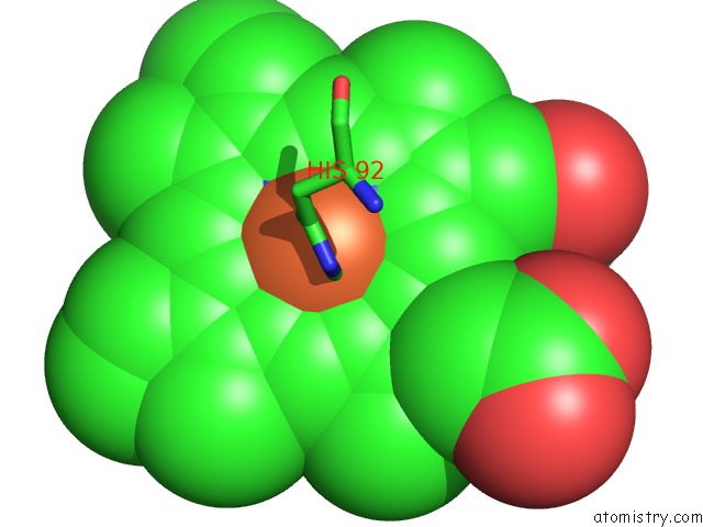
Mono view
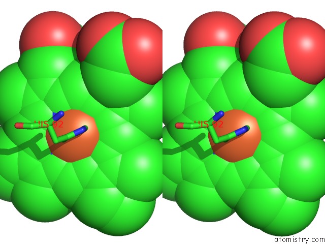
Stereo pair view
Mono view
Stereo pair view
A full contact list of Iron with other atoms in the Fe binding
site number 2 of Analysis of the Crystal Structure, Molecular Modeling and Infrared Spectroscopy of the Distal Beta-Heme Pocket VALINE67(E11)-Threonine Mutation of Hemoglobin within 5.0Å range:
|
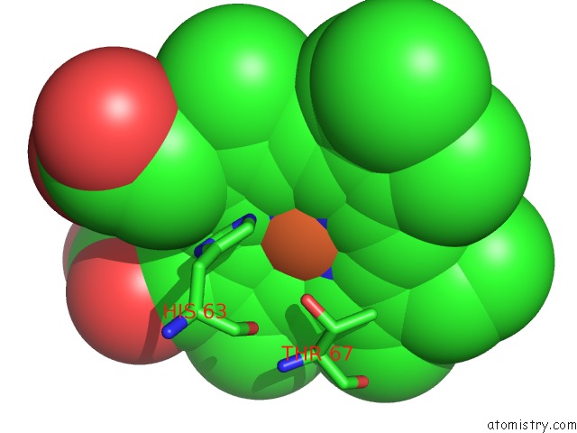
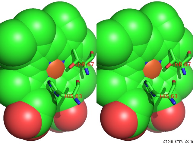
Iron binding site 3 out of 4 in 1hdb
Go back to
Iron binding site 3 out
of 4 in the Analysis of the Crystal Structure, Molecular Modeling and Infrared Spectroscopy of the Distal Beta-Heme Pocket VALINE67(E11)-Threonine Mutation of Hemoglobin
Mono view
Stereo pair view
Mono view
Stereo pair view
A full contact list of Iron with other atoms in the Fe binding
site number 3 of Analysis of the Crystal Structure, Molecular Modeling and Infrared Spectroscopy of the Distal Beta-Heme Pocket VALINE67(E11)-Threonine Mutation of Hemoglobin within 5.0Å range:
|
Iron binding site 4 out of 4 in 1hdb
Go back to
Iron binding site 4 out
of 4 in the Analysis of the Crystal Structure, Molecular Modeling and Infrared Spectroscopy of the Distal Beta-Heme Pocket VALINE67(E11)-Threonine Mutation of Hemoglobin
Mono view
Stereo pair view
Mono view
Stereo pair view
A full contact list of Iron with other atoms in the Fe binding
site number 4 of Analysis of the Crystal Structure, Molecular Modeling and Infrared Spectroscopy of the Distal Beta-Heme Pocket VALINE67(E11)-Threonine Mutation of Hemoglobin within 5.0Å range:
|
Reference:
I.Pechik,
X.Ji,
K.Fidelis,
M.Karavitis,
J.Moult,
W.S.Brinigar,
C.Fronticelli,
G.L.Gilliland.
Crystallographic, Molecular Modeling, and Biophysical Characterization of the Valine Beta 67 (E11)-->Threonine Variant of Hemoglobin. Biochemistry V. 35 1935 1996.
ISSN: ISSN 0006-2960
PubMed: 8639677
DOI: 10.1021/BI9519967
Page generated: Wed Jul 16 15:54:00 2025
ISSN: ISSN 0006-2960
PubMed: 8639677
DOI: 10.1021/BI9519967
Last articles
F in 9VCLF in 9VCK
F in 9JHS
F in 9JF9
F in 9CPK
Cu in 9OH6
Cu in 9OH7
Cu in 9VQM
Cl in 9QM5
Cl in 9RM2