Iron »
PDB 1hds-1hxq »
1hgb »
Iron in PDB 1hgb: High Resolution Crystal Structures and Comparisons of T State Deoxyhaemoglobin and Two Liganded T-State Haemoglobins: T(Alpha-Oxy) Haemoglobin and T(Met)Haemoglobin
Protein crystallography data
The structure of High Resolution Crystal Structures and Comparisons of T State Deoxyhaemoglobin and Two Liganded T-State Haemoglobins: T(Alpha-Oxy) Haemoglobin and T(Met)Haemoglobin, PDB code: 1hgb
was solved by
R.Liddington,
Z.Derewenda,
E.Dodson,
R.Hubbard,
G.Dodson,
with X-Ray Crystallography technique. A brief refinement statistics is given in the table below:
| Resolution Low / High (Å) | N/A / 2.10 |
| Space group | P 21 21 2 |
| Cell size a, b, c (Å), α, β, γ (°) | 95.780, 97.780, 65.490, 90.00, 90.00, 90.00 |
| R / Rfree (%) | n/a / n/a |
Iron Binding Sites:
The binding sites of Iron atom in the High Resolution Crystal Structures and Comparisons of T State Deoxyhaemoglobin and Two Liganded T-State Haemoglobins: T(Alpha-Oxy) Haemoglobin and T(Met)Haemoglobin
(pdb code 1hgb). This binding sites where shown within
5.0 Angstroms radius around Iron atom.
In total 4 binding sites of Iron where determined in the High Resolution Crystal Structures and Comparisons of T State Deoxyhaemoglobin and Two Liganded T-State Haemoglobins: T(Alpha-Oxy) Haemoglobin and T(Met)Haemoglobin, PDB code: 1hgb:
Jump to Iron binding site number: 1; 2; 3; 4;
In total 4 binding sites of Iron where determined in the High Resolution Crystal Structures and Comparisons of T State Deoxyhaemoglobin and Two Liganded T-State Haemoglobins: T(Alpha-Oxy) Haemoglobin and T(Met)Haemoglobin, PDB code: 1hgb:
Jump to Iron binding site number: 1; 2; 3; 4;
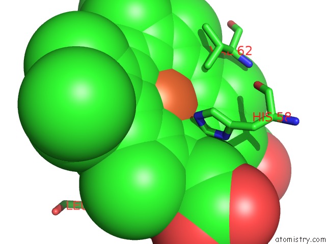
Iron binding site 1 out of 4 in 1hgb
Go back to
Iron binding site 1 out
of 4 in the High Resolution Crystal Structures and Comparisons of T State Deoxyhaemoglobin and Two Liganded T-State Haemoglobins: T(Alpha-Oxy) Haemoglobin and T(Met)Haemoglobin
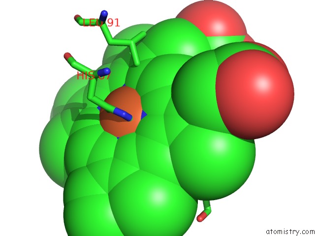
Mono view
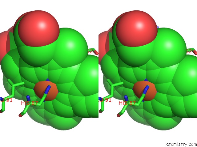
Stereo pair view
Mono view
Stereo pair view
A full contact list of Iron with other atoms in the Fe binding
site number 1 of High Resolution Crystal Structures and Comparisons of T State Deoxyhaemoglobin and Two Liganded T-State Haemoglobins: T(Alpha-Oxy) Haemoglobin and T(Met)Haemoglobin within 5.0Å range:
|
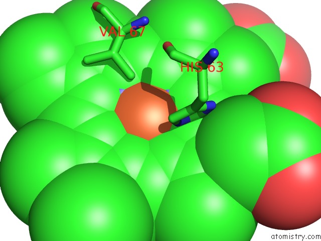
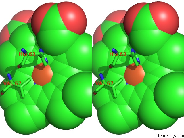
Iron binding site 2 out of 4 in 1hgb
Go back to
Iron binding site 2 out
of 4 in the High Resolution Crystal Structures and Comparisons of T State Deoxyhaemoglobin and Two Liganded T-State Haemoglobins: T(Alpha-Oxy) Haemoglobin and T(Met)Haemoglobin
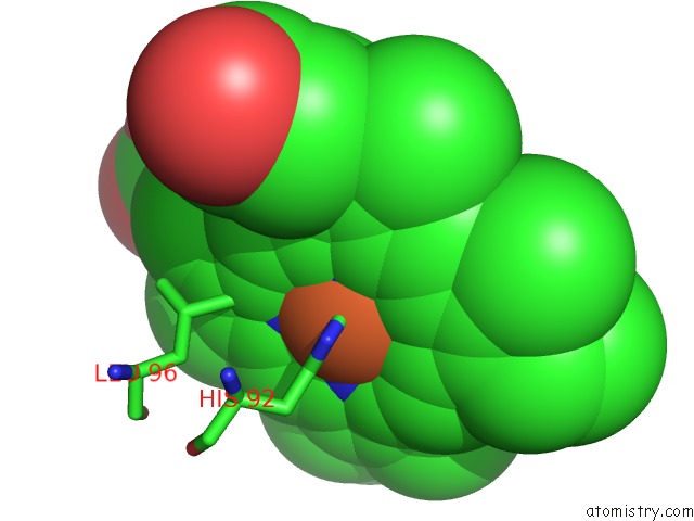
Mono view
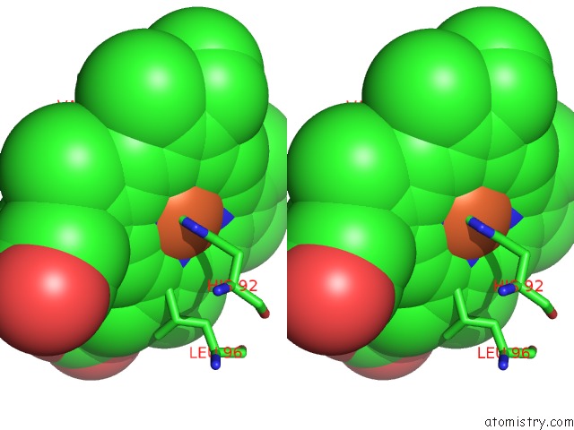
Stereo pair view
Mono view
Stereo pair view
A full contact list of Iron with other atoms in the Fe binding
site number 2 of High Resolution Crystal Structures and Comparisons of T State Deoxyhaemoglobin and Two Liganded T-State Haemoglobins: T(Alpha-Oxy) Haemoglobin and T(Met)Haemoglobin within 5.0Å range:
|
Iron binding site 3 out of 4 in 1hgb
Go back to
Iron binding site 3 out
of 4 in the High Resolution Crystal Structures and Comparisons of T State Deoxyhaemoglobin and Two Liganded T-State Haemoglobins: T(Alpha-Oxy) Haemoglobin and T(Met)Haemoglobin
Mono view

Stereo pair view
Mono view
Stereo pair view
A full contact list of Iron with other atoms in the Fe binding
site number 3 of High Resolution Crystal Structures and Comparisons of T State Deoxyhaemoglobin and Two Liganded T-State Haemoglobins: T(Alpha-Oxy) Haemoglobin and T(Met)Haemoglobin within 5.0Å range:
|
Iron binding site 4 out of 4 in 1hgb
Go back to
Iron binding site 4 out
of 4 in the High Resolution Crystal Structures and Comparisons of T State Deoxyhaemoglobin and Two Liganded T-State Haemoglobins: T(Alpha-Oxy) Haemoglobin and T(Met)Haemoglobin
Mono view
Stereo pair view
Mono view
Stereo pair view
A full contact list of Iron with other atoms in the Fe binding
site number 4 of High Resolution Crystal Structures and Comparisons of T State Deoxyhaemoglobin and Two Liganded T-State Haemoglobins: T(Alpha-Oxy) Haemoglobin and T(Met)Haemoglobin within 5.0Å range:
|
Reference:
R.Liddington,
Z.Derewenda,
E.Dodson,
R.Hubbard,
G.Dodson.
High Resolution Crystal Structures and Comparisons of T-State Deoxyhaemoglobin and Two Liganded T-State Haemoglobins: T(Alpha-Oxy)Haemoglobin and T(Met)Haemoglobin. J.Mol.Biol. V. 228 551 1992.
ISSN: ISSN 0022-2836
PubMed: 1453464
DOI: 10.1016/0022-2836(92)90842-8
Page generated: Wed Jul 16 15:57:52 2025
ISSN: ISSN 0022-2836
PubMed: 1453464
DOI: 10.1016/0022-2836(92)90842-8
Last articles
Zn in 1WGEZn in 1WFZ
Zn in 1WFP
Zn in 1WFL
Zn in 1WFX
Zn in 1WFK
Zn in 1WFF
Zn in 1WFH
Zn in 1WFE
Zn in 1WAA