Iron »
PDB 1hds-1hxq »
1hh7 »
Iron in PDB 1hh7: Refined Crystal Structure of Cytochrome C2 From Rhodopseudomonas Palustris at 1.4 Angstrom Resolution
Protein crystallography data
The structure of Refined Crystal Structure of Cytochrome C2 From Rhodopseudomonas Palustris at 1.4 Angstrom Resolution, PDB code: 1hh7
was solved by
G.Garau,
S.Geremia,
with X-Ray Crystallography technique. A brief refinement statistics is given in the table below:
| Resolution Low / High (Å) | 56.00 / 1.40 |
| Space group | P 32 2 1 |
| Cell size a, b, c (Å), α, β, γ (°) | 64.670, 64.670, 68.250, 90.00, 90.00, 120.00 |
| R / Rfree (%) | 15.1 / n/a |
Iron Binding Sites:
The binding sites of Iron atom in the Refined Crystal Structure of Cytochrome C2 From Rhodopseudomonas Palustris at 1.4 Angstrom Resolution
(pdb code 1hh7). This binding sites where shown within
5.0 Angstroms radius around Iron atom.
In total only one binding site of Iron was determined in the Refined Crystal Structure of Cytochrome C2 From Rhodopseudomonas Palustris at 1.4 Angstrom Resolution, PDB code: 1hh7:
In total only one binding site of Iron was determined in the Refined Crystal Structure of Cytochrome C2 From Rhodopseudomonas Palustris at 1.4 Angstrom Resolution, PDB code: 1hh7:
Iron binding site 1 out of 1 in 1hh7
Go back to
Iron binding site 1 out
of 1 in the Refined Crystal Structure of Cytochrome C2 From Rhodopseudomonas Palustris at 1.4 Angstrom Resolution
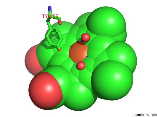
Mono view
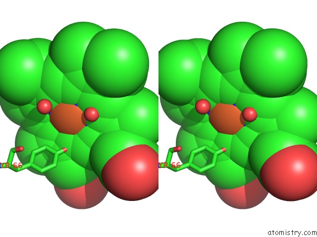
Stereo pair view
Mono view
Stereo pair view
A full contact list of Iron with other atoms in the Fe binding
site number 1 of Refined Crystal Structure of Cytochrome C2 From Rhodopseudomonas Palustris at 1.4 Angstrom Resolution within 5.0Å range:
|
Reference:
G.Garau,
S.Geremia,
L.Randaccio,
L.Vaccari,
M.S.Viezzoli.
Crystallization and Preliminary X-Ray Analysis of Two pH-Dependent Forms of Cytochrome C2 From Rhodopseudomonas Palustris Acta Crystallogr.,Sect.D V. 56 1699 2000.
ISSN: ISSN 0907-4449
PubMed: 11092951
DOI: 10.1107/S0907444900013573
Page generated: Wed Jul 16 15:59:09 2025
ISSN: ISSN 0907-4449
PubMed: 11092951
DOI: 10.1107/S0907444900013573
Last articles
Xe in 1URYXe in 1VAU
Xe in 1UVX
Xe in 1S56
Xe in 1UVY
Xe in 1UOC
Xe in 1UO6
Xe in 1U0X
Xe in 1KQN
Xe in 1RKY