Iron »
PDB 1hds-1hxq »
1ht8 »
Iron in PDB 1ht8: The 2.7 Angstrom Resolution Model of Ovine Cox-1 Complexed with Alclofenac
Enzymatic activity of The 2.7 Angstrom Resolution Model of Ovine Cox-1 Complexed with Alclofenac
All present enzymatic activity of The 2.7 Angstrom Resolution Model of Ovine Cox-1 Complexed with Alclofenac:
1.14.99.1;
1.14.99.1;
Protein crystallography data
The structure of The 2.7 Angstrom Resolution Model of Ovine Cox-1 Complexed with Alclofenac, PDB code: 1ht8
was solved by
B.S.Selinsky,
K.Gupta,
C.T.Sharkey,
P.J.Loll,
with X-Ray Crystallography technique. A brief refinement statistics is given in the table below:
| Resolution Low / High (Å) | 19.92 / 2.69 |
| Space group | I 2 2 2 |
| Cell size a, b, c (Å), α, β, γ (°) | 99.149, 208.450, 222.399, 90.00, 90.00, 90.00 |
| R / Rfree (%) | 21.1 / 24.2 |
Other elements in 1ht8:
The structure of The 2.7 Angstrom Resolution Model of Ovine Cox-1 Complexed with Alclofenac also contains other interesting chemical elements:
| Chlorine | (Cl) | 2 atoms |
Iron Binding Sites:
The binding sites of Iron atom in the The 2.7 Angstrom Resolution Model of Ovine Cox-1 Complexed with Alclofenac
(pdb code 1ht8). This binding sites where shown within
5.0 Angstroms radius around Iron atom.
In total 2 binding sites of Iron where determined in the The 2.7 Angstrom Resolution Model of Ovine Cox-1 Complexed with Alclofenac, PDB code: 1ht8:
Jump to Iron binding site number: 1; 2;
In total 2 binding sites of Iron where determined in the The 2.7 Angstrom Resolution Model of Ovine Cox-1 Complexed with Alclofenac, PDB code: 1ht8:
Jump to Iron binding site number: 1; 2;
Iron binding site 1 out of 2 in 1ht8
Go back to
Iron binding site 1 out
of 2 in the The 2.7 Angstrom Resolution Model of Ovine Cox-1 Complexed with Alclofenac
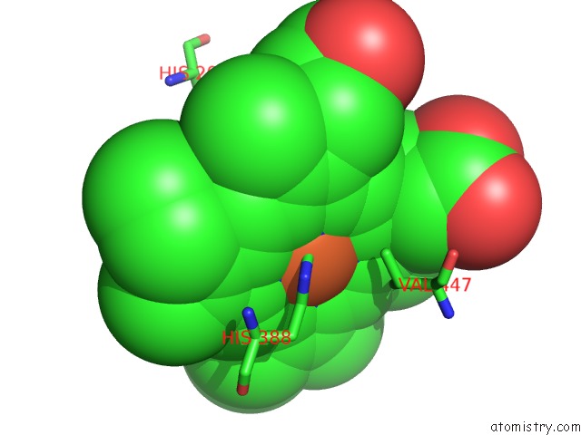
Mono view
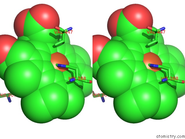
Stereo pair view
Mono view
Stereo pair view
A full contact list of Iron with other atoms in the Fe binding
site number 1 of The 2.7 Angstrom Resolution Model of Ovine Cox-1 Complexed with Alclofenac within 5.0Å range:
|
Iron binding site 2 out of 2 in 1ht8
Go back to
Iron binding site 2 out
of 2 in the The 2.7 Angstrom Resolution Model of Ovine Cox-1 Complexed with Alclofenac
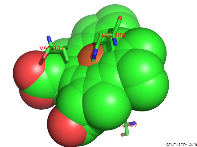
Mono view
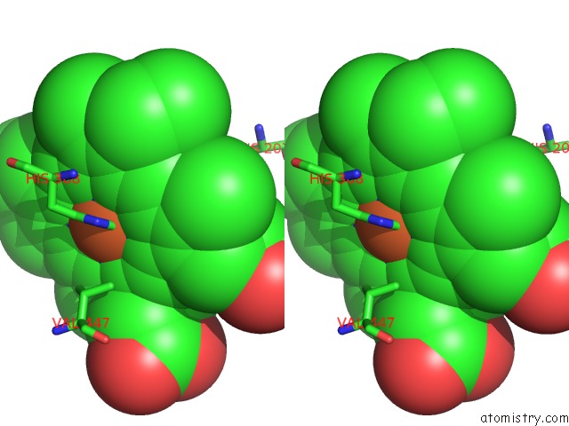
Stereo pair view
Mono view
Stereo pair view
A full contact list of Iron with other atoms in the Fe binding
site number 2 of The 2.7 Angstrom Resolution Model of Ovine Cox-1 Complexed with Alclofenac within 5.0Å range:
|
Reference:
B.S.Selinsky,
K.Gupta,
C.T.Sharkey,
P.J.Loll.
Structural Analysis of Nsaid Binding By Prostaglandin H2 Synthase: Time-Dependent and Time-Independent Inhibitors Elicit Identical Enzyme Conformations. Biochemistry V. 40 5172 2001.
ISSN: ISSN 0006-2960
PubMed: 11318639
DOI: 10.1021/BI010045S
Page generated: Wed Jul 16 16:07:53 2025
ISSN: ISSN 0006-2960
PubMed: 11318639
DOI: 10.1021/BI010045S
Last articles
K in 7POZK in 7PNL
K in 7POE
K in 7PLK
K in 7PKA
K in 7PHK
K in 7PHL
K in 7PJI
K in 7PHI
K in 7PHH