Iron »
PDB 1hds-1hxq »
1hv4 »
Iron in PDB 1hv4: Crystal Structure Analysis of Bar-Head Goose Hemoglobin (Deoxy Form)
Protein crystallography data
The structure of Crystal Structure Analysis of Bar-Head Goose Hemoglobin (Deoxy Form), PDB code: 1hv4
was solved by
Y.Liang,
Z.Hua,
X.Liang,
Q.Xu,
G.Lu,
with X-Ray Crystallography technique. A brief refinement statistics is given in the table below:
| Resolution Low / High (Å) | 36.89 / 2.80 |
| Space group | P 1 |
| Cell size a, b, c (Å), α, β, γ (°) | 70.660, 94.100, 59.230, 71.55, 65.10, 83.10 |
| R / Rfree (%) | 19.7 / 24.3 |
Iron Binding Sites:
The binding sites of Iron atom in the Crystal Structure Analysis of Bar-Head Goose Hemoglobin (Deoxy Form)
(pdb code 1hv4). This binding sites where shown within
5.0 Angstroms radius around Iron atom.
In total 8 binding sites of Iron where determined in the Crystal Structure Analysis of Bar-Head Goose Hemoglobin (Deoxy Form), PDB code: 1hv4:
Jump to Iron binding site number: 1; 2; 3; 4; 5; 6; 7; 8;
In total 8 binding sites of Iron where determined in the Crystal Structure Analysis of Bar-Head Goose Hemoglobin (Deoxy Form), PDB code: 1hv4:
Jump to Iron binding site number: 1; 2; 3; 4; 5; 6; 7; 8;
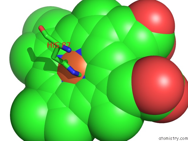
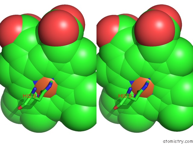
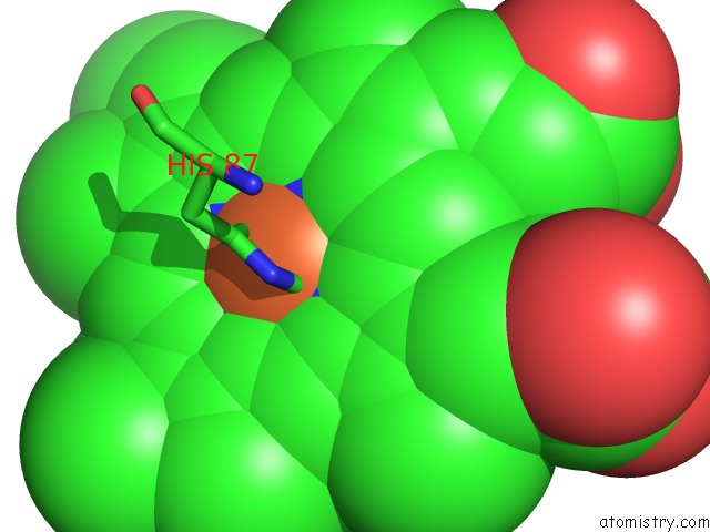
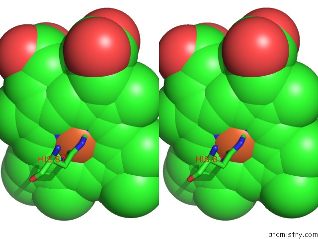
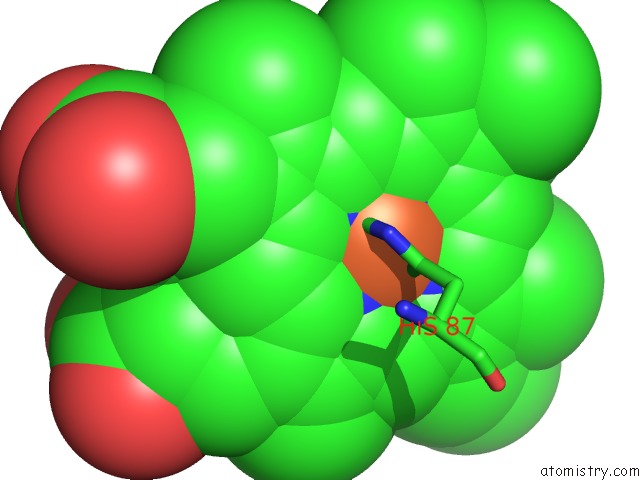
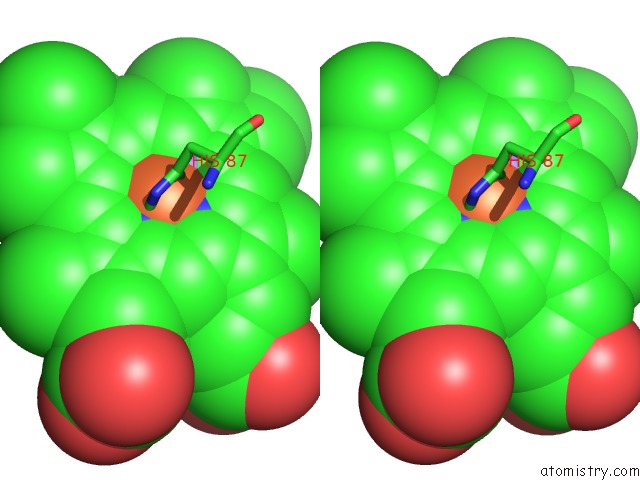
Iron binding site 1 out of 8 in 1hv4
Go back to
Iron binding site 1 out
of 8 in the Crystal Structure Analysis of Bar-Head Goose Hemoglobin (Deoxy Form)
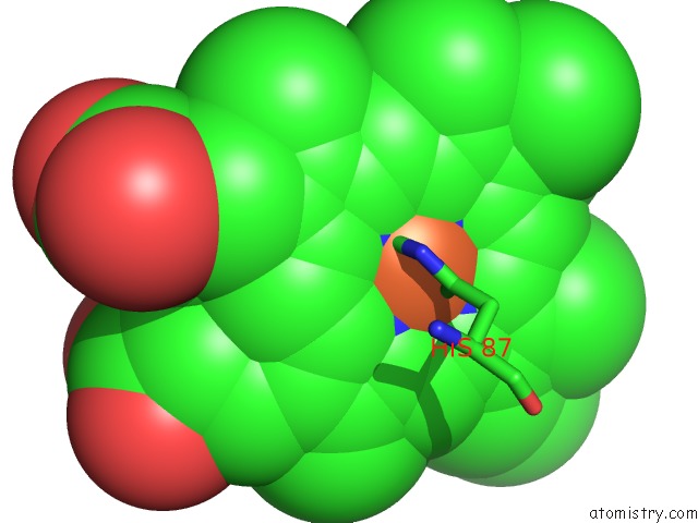
Mono view
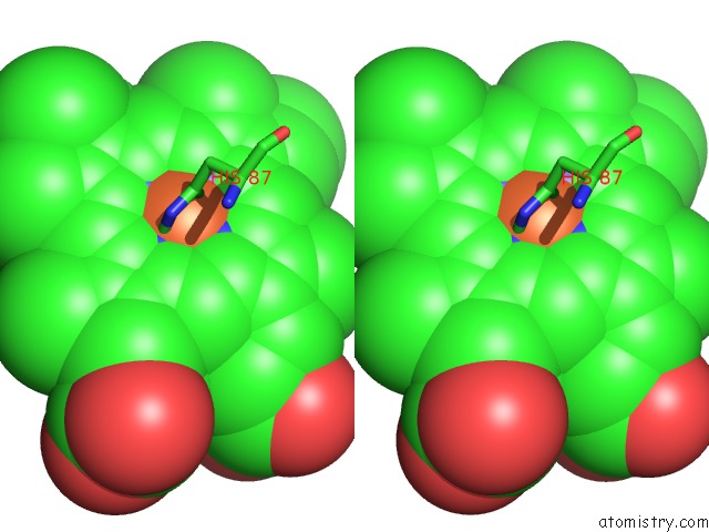
Stereo pair view
Mono view
Stereo pair view
A full contact list of Iron with other atoms in the Fe binding
site number 1 of Crystal Structure Analysis of Bar-Head Goose Hemoglobin (Deoxy Form) within 5.0Å range:
|
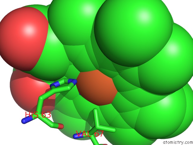
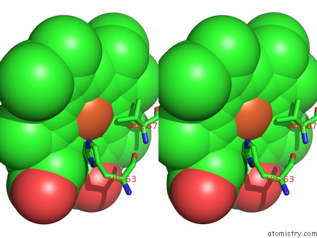
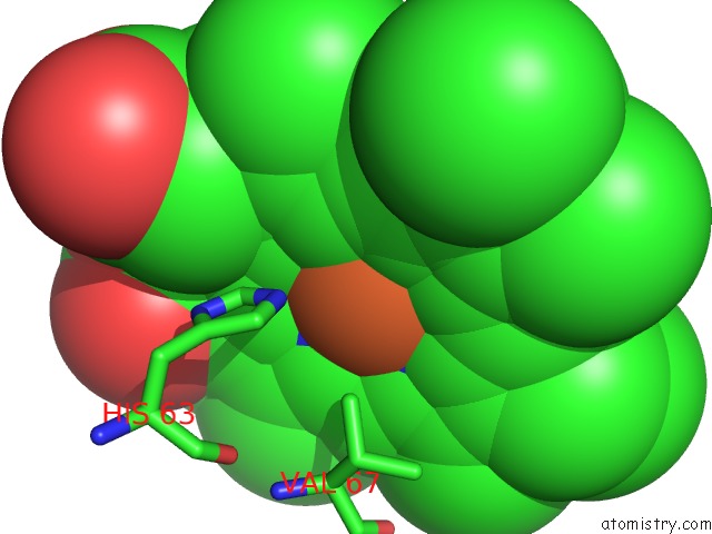
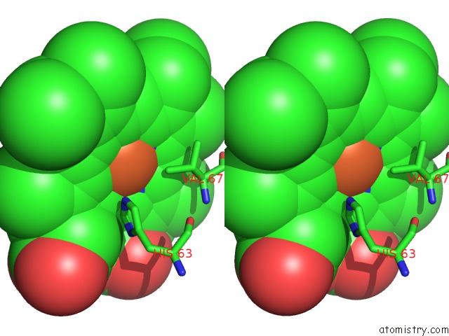
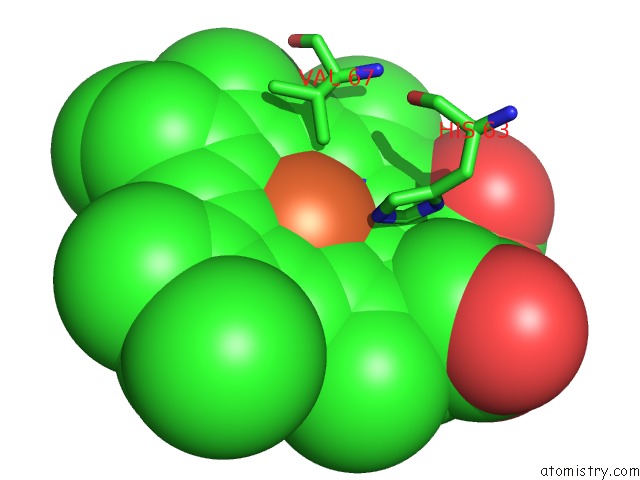
Iron binding site 2 out of 8 in 1hv4
Go back to
Iron binding site 2 out
of 8 in the Crystal Structure Analysis of Bar-Head Goose Hemoglobin (Deoxy Form)
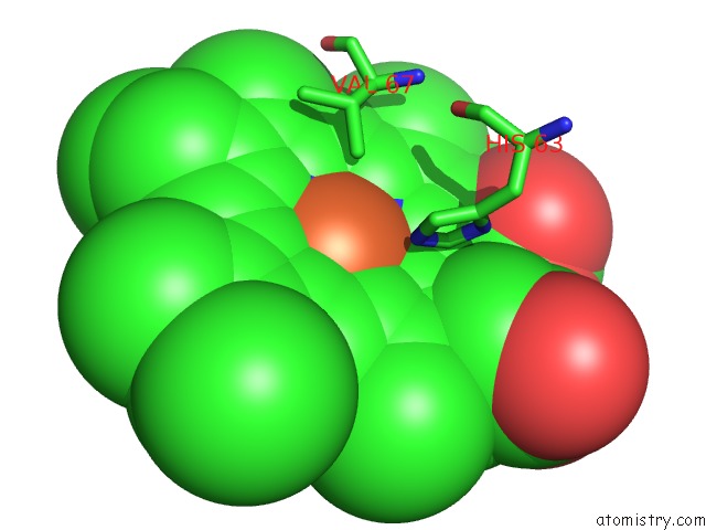
Mono view
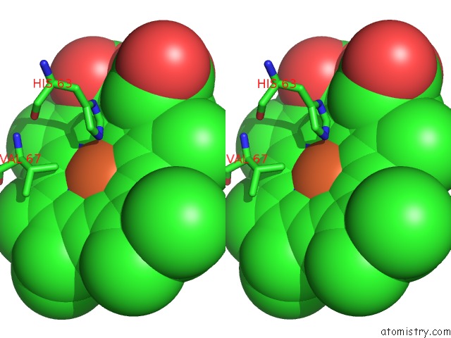
Stereo pair view
Mono view
Stereo pair view
A full contact list of Iron with other atoms in the Fe binding
site number 2 of Crystal Structure Analysis of Bar-Head Goose Hemoglobin (Deoxy Form) within 5.0Å range:
|
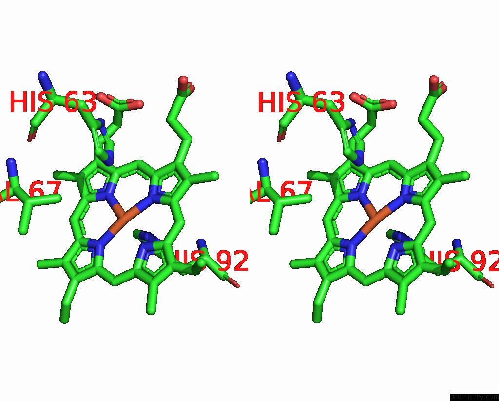
Iron binding site 3 out of 8 in 1hv4
Go back to
Iron binding site 3 out
of 8 in the Crystal Structure Analysis of Bar-Head Goose Hemoglobin (Deoxy Form)
Mono view
Stereo pair view
Mono view
Stereo pair view
A full contact list of Iron with other atoms in the Fe binding
site number 3 of Crystal Structure Analysis of Bar-Head Goose Hemoglobin (Deoxy Form) within 5.0Å range:
|
Iron binding site 4 out of 8 in 1hv4
Go back to
Iron binding site 4 out
of 8 in the Crystal Structure Analysis of Bar-Head Goose Hemoglobin (Deoxy Form)
Mono view
Stereo pair view
Mono view
Stereo pair view
A full contact list of Iron with other atoms in the Fe binding
site number 4 of Crystal Structure Analysis of Bar-Head Goose Hemoglobin (Deoxy Form) within 5.0Å range:
|
Iron binding site 5 out of 8 in 1hv4
Go back to
Iron binding site 5 out
of 8 in the Crystal Structure Analysis of Bar-Head Goose Hemoglobin (Deoxy Form)
Mono view
Stereo pair view
Mono view
Stereo pair view
A full contact list of Iron with other atoms in the Fe binding
site number 5 of Crystal Structure Analysis of Bar-Head Goose Hemoglobin (Deoxy Form) within 5.0Å range:
|
Iron binding site 6 out of 8 in 1hv4
Go back to
Iron binding site 6 out
of 8 in the Crystal Structure Analysis of Bar-Head Goose Hemoglobin (Deoxy Form)
Mono view
Stereo pair view
Mono view
Stereo pair view
A full contact list of Iron with other atoms in the Fe binding
site number 6 of Crystal Structure Analysis of Bar-Head Goose Hemoglobin (Deoxy Form) within 5.0Å range:
|
Iron binding site 7 out of 8 in 1hv4
Go back to
Iron binding site 7 out
of 8 in the Crystal Structure Analysis of Bar-Head Goose Hemoglobin (Deoxy Form)
Mono view
Stereo pair view
Mono view
Stereo pair view
A full contact list of Iron with other atoms in the Fe binding
site number 7 of Crystal Structure Analysis of Bar-Head Goose Hemoglobin (Deoxy Form) within 5.0Å range:
|
Iron binding site 8 out of 8 in 1hv4
Go back to
Iron binding site 8 out
of 8 in the Crystal Structure Analysis of Bar-Head Goose Hemoglobin (Deoxy Form)
Mono view
Stereo pair view
Mono view
Stereo pair view
A full contact list of Iron with other atoms in the Fe binding
site number 8 of Crystal Structure Analysis of Bar-Head Goose Hemoglobin (Deoxy Form) within 5.0Å range:
|
Reference:
Y.Liang,
Z.Hua,
X.Liang,
Q.Xu,
G.Lu.
The Crystal Structure of Bar-Headed Goose Hemoglobin in Deoxy Form: the Allosteric Mechanism of A Hemoglobin Species with High Oxygen Affinity. J.Mol.Biol. V. 313 123 2001.
ISSN: ISSN 0022-2836
PubMed: 11601851
DOI: 10.1006/JMBI.2001.5028
Page generated: Wed Jul 16 16:08:32 2025
ISSN: ISSN 0022-2836
PubMed: 11601851
DOI: 10.1006/JMBI.2001.5028
Last articles
W in 9FPPW in 8PRM
W in 9QM1
W in 9QM0
W in 9OJ3
W in 9MQX
W in 9FP4
W in 9BEO
W in 9BEM
W in 8P2U