Iron »
PDB 1hzu-1ird »
1iej »
Iron in PDB 1iej: Ovotransferrin, N-Terminal Lobe, Holo Form, at 1.65 A Resolution
Protein crystallography data
The structure of Ovotransferrin, N-Terminal Lobe, Holo Form, at 1.65 A Resolution, PDB code: 1iej
was solved by
K.Mizutani,
B.Mikami,
M.Hirose,
with X-Ray Crystallography technique. A brief refinement statistics is given in the table below:
| Resolution Low / High (Å) | 10.00 / 1.65 |
| Space group | P 21 21 21 |
| Cell size a, b, c (Å), α, β, γ (°) | 46.460, 85.950, 76.100, 90.00, 90.00, 90.00 |
| R / Rfree (%) | 17.3 / 24.2 |
Iron Binding Sites:
The binding sites of Iron atom in the Ovotransferrin, N-Terminal Lobe, Holo Form, at 1.65 A Resolution
(pdb code 1iej). This binding sites where shown within
5.0 Angstroms radius around Iron atom.
In total only one binding site of Iron was determined in the Ovotransferrin, N-Terminal Lobe, Holo Form, at 1.65 A Resolution, PDB code: 1iej:
In total only one binding site of Iron was determined in the Ovotransferrin, N-Terminal Lobe, Holo Form, at 1.65 A Resolution, PDB code: 1iej:
Iron binding site 1 out of 1 in 1iej
Go back to
Iron binding site 1 out
of 1 in the Ovotransferrin, N-Terminal Lobe, Holo Form, at 1.65 A Resolution
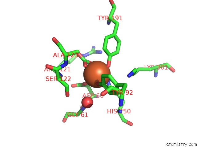
Mono view
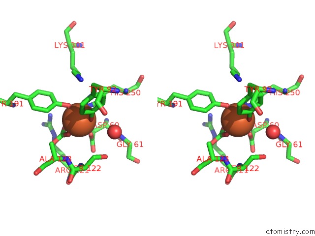
Stereo pair view
Mono view
Stereo pair view
A full contact list of Iron with other atoms in the Fe binding
site number 1 of Ovotransferrin, N-Terminal Lobe, Holo Form, at 1.65 A Resolution within 5.0Å range:
|
Reference:
K.Mizutani,
B.Mikami,
M.Hirose.
Domain Closure Mechanism in Transferrins: New Viewpoints About the Hinge Structure and Motion As Deduced From High Resolution Crystal Structures of Ovotransferrin N-Lobe. J.Mol.Biol. V. 309 937 2001.
ISSN: ISSN 0022-2836
PubMed: 11399070
DOI: 10.1006/JMBI.2001.4719
Page generated: Wed Jul 16 16:15:20 2025
ISSN: ISSN 0022-2836
PubMed: 11399070
DOI: 10.1006/JMBI.2001.4719
Last articles
Xe in 2DKIXe in 2FIC
Xe in 2A7A
Xe in 2A7D
Xe in 2A9R
Xe in 2A7B
Xe in 1ZDM
Xe in 2A7C
Xe in 1W53
Xe in 1W2Z