Iron »
PDB 1hzu-1ird »
1ikj »
Iron in PDB 1ikj: 1.27 A Crystal Structure of Nitrophorin 4 From Rhodnius Prolixus Complexed with Imidazole
Protein crystallography data
The structure of 1.27 A Crystal Structure of Nitrophorin 4 From Rhodnius Prolixus Complexed with Imidazole, PDB code: 1ikj
was solved by
S.A.Roberts,
A.Weichsel,
Y.Qui,
J.A.Shelnutt,
F.A.Walker,
W.R.Montfort,
with X-Ray Crystallography technique. A brief refinement statistics is given in the table below:
| Resolution Low / High (Å) | 22.00 / 1.27 |
| Space group | C 1 2 1 |
| Cell size a, b, c (Å), α, β, γ (°) | 70.180, 42.480, 52.960, 90.00, 94.28, 90.00 |
| R / Rfree (%) | 13 / 17 |
Iron Binding Sites:
The binding sites of Iron atom in the 1.27 A Crystal Structure of Nitrophorin 4 From Rhodnius Prolixus Complexed with Imidazole
(pdb code 1ikj). This binding sites where shown within
5.0 Angstroms radius around Iron atom.
In total only one binding site of Iron was determined in the 1.27 A Crystal Structure of Nitrophorin 4 From Rhodnius Prolixus Complexed with Imidazole, PDB code: 1ikj:
In total only one binding site of Iron was determined in the 1.27 A Crystal Structure of Nitrophorin 4 From Rhodnius Prolixus Complexed with Imidazole, PDB code: 1ikj:
Iron binding site 1 out of 1 in 1ikj
Go back to
Iron binding site 1 out
of 1 in the 1.27 A Crystal Structure of Nitrophorin 4 From Rhodnius Prolixus Complexed with Imidazole
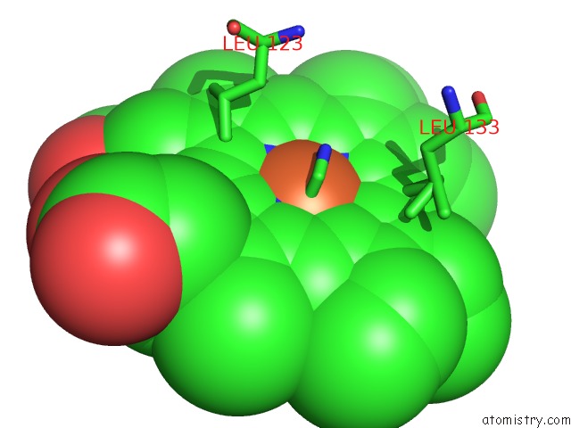
Mono view
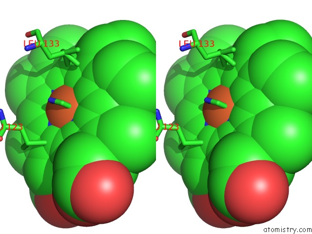
Stereo pair view
Mono view
Stereo pair view
A full contact list of Iron with other atoms in the Fe binding
site number 1 of 1.27 A Crystal Structure of Nitrophorin 4 From Rhodnius Prolixus Complexed with Imidazole within 5.0Å range:
|
Reference:
S.A.Roberts,
A.Weichsel,
Y.Qiu,
J.A.Shelnutt,
F.A.Walker,
W.R.Montfort.
Ligand-Induced Heme Ruffling and Bent No Geometry in Ultra-High-Resolution Structures of Nitrophorin 4. Biochemistry V. 40 11327 2001.
ISSN: ISSN 0006-2960
PubMed: 11560480
DOI: 10.1021/BI0109257
Page generated: Wed Jul 16 16:15:44 2025
ISSN: ISSN 0006-2960
PubMed: 11560480
DOI: 10.1021/BI0109257
Last articles
Zn in 1SHNZn in 1SLX
Zn in 1SFO
Zn in 1SKU
Zn in 1SHQ
Zn in 1SHW
Zn in 1SGF
Zn in 1SG0
Zn in 1SG6
Zn in 1SED