Iron »
PDB 1hzu-1ird »
1io3 »
Iron in PDB 1io3: Crystal Structure of Ferricytochrome C2 From Rhodopseudomonas Viridis
Protein crystallography data
The structure of Crystal Structure of Ferricytochrome C2 From Rhodopseudomonas Viridis, PDB code: 1io3
was solved by
K.Miki,
S.Sogabe,
with X-Ray Crystallography technique. A brief refinement statistics is given in the table below:
| Resolution Low / High (Å) | 20.00 / 1.90 |
| Space group | P 32 2 1 |
| Cell size a, b, c (Å), α, β, γ (°) | 75.970, 75.970, 40.370, 90.00, 90.00, 120.00 |
| R / Rfree (%) | 20.8 / 24.3 |
Iron Binding Sites:
The binding sites of Iron atom in the Crystal Structure of Ferricytochrome C2 From Rhodopseudomonas Viridis
(pdb code 1io3). This binding sites where shown within
5.0 Angstroms radius around Iron atom.
In total only one binding site of Iron was determined in the Crystal Structure of Ferricytochrome C2 From Rhodopseudomonas Viridis, PDB code: 1io3:
In total only one binding site of Iron was determined in the Crystal Structure of Ferricytochrome C2 From Rhodopseudomonas Viridis, PDB code: 1io3:
Iron binding site 1 out of 1 in 1io3
Go back to
Iron binding site 1 out
of 1 in the Crystal Structure of Ferricytochrome C2 From Rhodopseudomonas Viridis
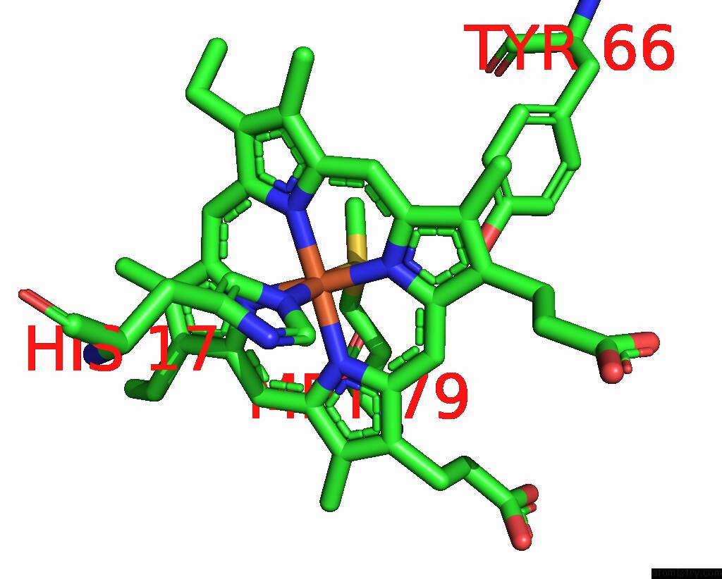
Mono view
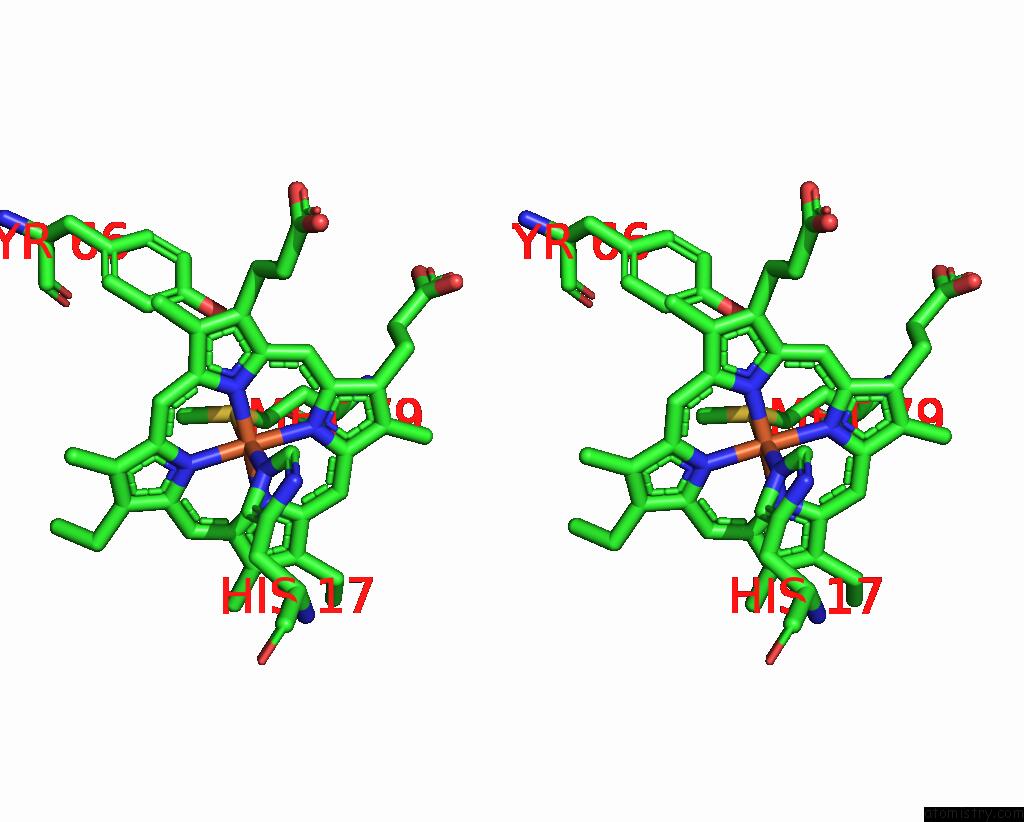
Stereo pair view
Mono view
Stereo pair view
A full contact list of Iron with other atoms in the Fe binding
site number 1 of Crystal Structure of Ferricytochrome C2 From Rhodopseudomonas Viridis within 5.0Å range:
|
Reference:
S.Sogabe,
K.Miki.
Crystal Structure of the Oxidized Cytochrome C(2) From Blastochloris Viridis. Febs Lett. V. 491 174 2001.
ISSN: ISSN 0014-5793
PubMed: 11240122
DOI: 10.1016/S0014-5793(01)02179-2
Page generated: Wed Jul 16 16:16:11 2025
ISSN: ISSN 0014-5793
PubMed: 11240122
DOI: 10.1016/S0014-5793(01)02179-2
Last articles
Zn in 1IZBZn in 1IY7
Zn in 1IX1
Zn in 1IWL
Zn in 1IS8
Zn in 1IUJ
Zn in 1ITU
Zn in 1ITQ
Zn in 1IRX
Zn in 1IT8