Iron »
PDB 1hzu-1ird »
1iop »
Iron in PDB 1iop: Incorporation of A Hemin with the Shortest Acid Side-Chains Into Myoglobin
Protein crystallography data
The structure of Incorporation of A Hemin with the Shortest Acid Side-Chains Into Myoglobin, PDB code: 1iop
was solved by
N.Igarashi,
S.Neya,
N.Funasaki,
N.Tanaka,
with X-Ray Crystallography technique. A brief refinement statistics is given in the table below:
| Resolution Low / High (Å) | 10.00 / 1.90 |
| Space group | P 6 |
| Cell size a, b, c (Å), α, β, γ (°) | 91.020, 91.020, 45.680, 90.00, 90.00, 120.00 |
| R / Rfree (%) | n/a / n/a |
Iron Binding Sites:
The binding sites of Iron atom in the Incorporation of A Hemin with the Shortest Acid Side-Chains Into Myoglobin
(pdb code 1iop). This binding sites where shown within
5.0 Angstroms radius around Iron atom.
In total only one binding site of Iron was determined in the Incorporation of A Hemin with the Shortest Acid Side-Chains Into Myoglobin, PDB code: 1iop:
In total only one binding site of Iron was determined in the Incorporation of A Hemin with the Shortest Acid Side-Chains Into Myoglobin, PDB code: 1iop:
Iron binding site 1 out of 1 in 1iop
Go back to
Iron binding site 1 out
of 1 in the Incorporation of A Hemin with the Shortest Acid Side-Chains Into Myoglobin
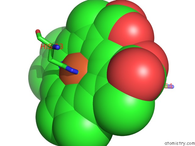
Mono view
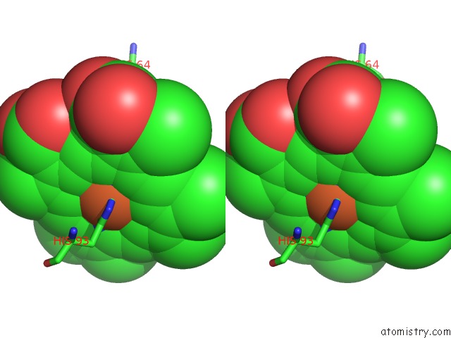
Stereo pair view
Mono view
Stereo pair view
A full contact list of Iron with other atoms in the Fe binding
site number 1 of Incorporation of A Hemin with the Shortest Acid Side-Chains Into Myoglobin within 5.0Å range:
|
Reference:
S.Neya,
N.Funasaki,
N.Igarashi,
A.Ikezaki,
T.Sato,
K.Imai,
N.Tanaka.
Structure and Function of 6,7-Dicarboxyheme-Substituted Myoglobin Biochemistry V. 37 5487 1998.
ISSN: ISSN 0006-2960
PubMed: 9548931
DOI: 10.1021/BI972632C
Page generated: Wed Jul 16 16:17:05 2025
ISSN: ISSN 0006-2960
PubMed: 9548931
DOI: 10.1021/BI972632C
Last articles
W in 9FPPW in 8PRM
W in 9QM1
W in 9QM0
W in 9OJ3
W in 9MQX
W in 9FP4
W in 9BEO
W in 9BEM
W in 8P2U