Iron »
PDB 1iro-1j3y »
1iu5 »
Iron in PDB 1iu5: X-Ray Crystal Structure of the Rubredoxin Mutant From Pyrococcus Furiosus
Protein crystallography data
The structure of X-Ray Crystal Structure of the Rubredoxin Mutant From Pyrococcus Furiosus, PDB code: 1iu5
was solved by
T.Chatake,
K.Kurihara,
I.Tanaka,
I.Tsyba,
R.Bau,
F.E.Jenney,
M.W.W.Adams,
N.Niimura,
with X-Ray Crystallography technique. A brief refinement statistics is given in the table below:
| Resolution Low / High (Å) | 10.00 / 1.50 |
| Space group | P 21 21 21 |
| Cell size a, b, c (Å), α, β, γ (°) | 34.480, 35.700, 43.160, 90.00, 90.00, 90.00 |
| R / Rfree (%) | 18.7 / 20.3 |
Iron Binding Sites:
The binding sites of Iron atom in the X-Ray Crystal Structure of the Rubredoxin Mutant From Pyrococcus Furiosus
(pdb code 1iu5). This binding sites where shown within
5.0 Angstroms radius around Iron atom.
In total only one binding site of Iron was determined in the X-Ray Crystal Structure of the Rubredoxin Mutant From Pyrococcus Furiosus, PDB code: 1iu5:
In total only one binding site of Iron was determined in the X-Ray Crystal Structure of the Rubredoxin Mutant From Pyrococcus Furiosus, PDB code: 1iu5:
Iron binding site 1 out of 1 in 1iu5
Go back to
Iron binding site 1 out
of 1 in the X-Ray Crystal Structure of the Rubredoxin Mutant From Pyrococcus Furiosus
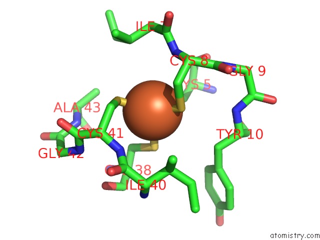
Mono view
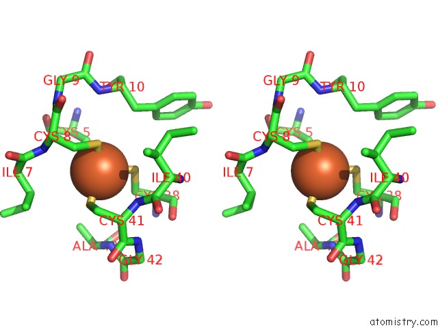
Stereo pair view
Mono view
Stereo pair view
A full contact list of Iron with other atoms in the Fe binding
site number 1 of X-Ray Crystal Structure of the Rubredoxin Mutant From Pyrococcus Furiosus within 5.0Å range:
|
Reference:
T.Chatake,
K.Kurihara,
I.Tanaka,
I.Tsyba,
R.Bau,
F.E.Jenney,
M.W.Adams,
N.Niimura.
A Neutron Crystallographic Analysis of A Rubredoxin Mutant at 1.6 A Resolution. Acta Crystallogr.,Sect.D V. 60 1364 2004.
ISSN: ISSN 0907-4449
PubMed: 15272158
DOI: 10.1107/S090744490401176X
Page generated: Wed Jul 16 16:21:32 2025
ISSN: ISSN 0907-4449
PubMed: 15272158
DOI: 10.1107/S090744490401176X
Last articles
Fe in 2YXOFe in 2YRS
Fe in 2YXC
Fe in 2YNM
Fe in 2YVJ
Fe in 2YP1
Fe in 2YU2
Fe in 2YU1
Fe in 2YQB
Fe in 2YOO