Iron »
PDB 1jl6-1k2o »
1jm1 »
Iron in PDB 1jm1: Crystal Structure of the Soluble Domain of the Rieske Protein II (Soxf) From Sulfolobus Acidocaldarius
Protein crystallography data
The structure of Crystal Structure of the Soluble Domain of the Rieske Protein II (Soxf) From Sulfolobus Acidocaldarius, PDB code: 1jm1
was solved by
H.Boenisch,
C.L.Schmidt,
G.Schaefer,
R.Ladenstein,
with X-Ray Crystallography technique. A brief refinement statistics is given in the table below:
| Resolution Low / High (Å) | 20.00 / 1.11 |
| Space group | P 61 |
| Cell size a, b, c (Å), α, β, γ (°) | 80.252, 80.252, 75.624, 90.00, 90.00, 120.00 |
| R / Rfree (%) | 10.6 / 12.5 |
Other elements in 1jm1:
The structure of Crystal Structure of the Soluble Domain of the Rieske Protein II (Soxf) From Sulfolobus Acidocaldarius also contains other interesting chemical elements:
| Magnesium | (Mg) | 1 atom |
Iron Binding Sites:
The binding sites of Iron atom in the Crystal Structure of the Soluble Domain of the Rieske Protein II (Soxf) From Sulfolobus Acidocaldarius
(pdb code 1jm1). This binding sites where shown within
5.0 Angstroms radius around Iron atom.
In total 2 binding sites of Iron where determined in the Crystal Structure of the Soluble Domain of the Rieske Protein II (Soxf) From Sulfolobus Acidocaldarius, PDB code: 1jm1:
Jump to Iron binding site number: 1; 2;
In total 2 binding sites of Iron where determined in the Crystal Structure of the Soluble Domain of the Rieske Protein II (Soxf) From Sulfolobus Acidocaldarius, PDB code: 1jm1:
Jump to Iron binding site number: 1; 2;
Iron binding site 1 out of 2 in 1jm1
Go back to
Iron binding site 1 out
of 2 in the Crystal Structure of the Soluble Domain of the Rieske Protein II (Soxf) From Sulfolobus Acidocaldarius
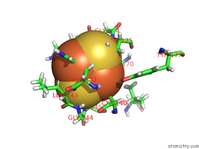
Mono view
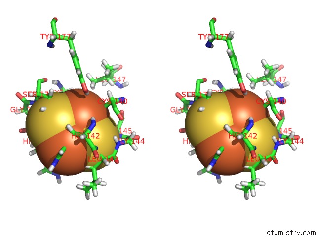
Stereo pair view
Mono view
Stereo pair view
A full contact list of Iron with other atoms in the Fe binding
site number 1 of Crystal Structure of the Soluble Domain of the Rieske Protein II (Soxf) From Sulfolobus Acidocaldarius within 5.0Å range:
|
Iron binding site 2 out of 2 in 1jm1
Go back to
Iron binding site 2 out
of 2 in the Crystal Structure of the Soluble Domain of the Rieske Protein II (Soxf) From Sulfolobus Acidocaldarius
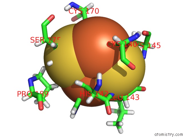
Mono view
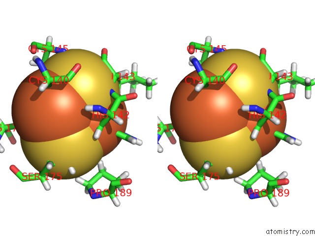
Stereo pair view
Mono view
Stereo pair view
A full contact list of Iron with other atoms in the Fe binding
site number 2 of Crystal Structure of the Soluble Domain of the Rieske Protein II (Soxf) From Sulfolobus Acidocaldarius within 5.0Å range:
|
Reference:
H.Bonisch,
C.L.Schmidt,
G.Schafer,
R.Ladenstein.
The Structure of the Soluble Domain of An Archaeal Rieske Iron-Sulfur Protein at 1.1 A Resolution. J.Mol.Biol. V. 319 791 2002.
ISSN: ISSN 0022-2836
PubMed: 12054871
DOI: 10.1016/S0022-2836(02)00323-6
Page generated: Wed Jul 16 16:35:11 2025
ISSN: ISSN 0022-2836
PubMed: 12054871
DOI: 10.1016/S0022-2836(02)00323-6
Last articles
K in 1J5YK in 1IS8
K in 1IS7
K in 1IWB
K in 1J51
K in 1IWP
K in 1I0P
K in 1IJV
K in 1HQ1
K in 1IH7