Iron »
PDB 1k2r-1kqg »
1k2v »
Iron in PDB 1k2v: E. Coli Periplasmic Protein Fhud Complexed with Desferal
Protein crystallography data
The structure of E. Coli Periplasmic Protein Fhud Complexed with Desferal, PDB code: 1k2v
was solved by
T.E.Clarke,
V.Braun,
G.Winkelmann,
L.W.Tari,
H.J.Vogel,
with X-Ray Crystallography technique. A brief refinement statistics is given in the table below:
| Resolution Low / High (Å) | 30.00 / 1.97 |
| Space group | P 63 |
| Cell size a, b, c (Å), α, β, γ (°) | 85.570, 85.570, 91.600, 90.00, 90.00, 120.00 |
| R / Rfree (%) | 22 / 24.1 |
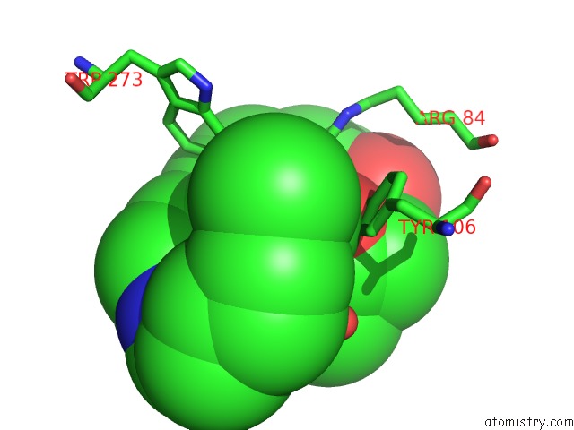
Iron Binding Sites:
The binding sites of Iron atom in the E. Coli Periplasmic Protein Fhud Complexed with Desferal
(pdb code 1k2v). This binding sites where shown within
5.0 Angstroms radius around Iron atom.
In total only one binding site of Iron was determined in the E. Coli Periplasmic Protein Fhud Complexed with Desferal, PDB code: 1k2v:
In total only one binding site of Iron was determined in the E. Coli Periplasmic Protein Fhud Complexed with Desferal, PDB code: 1k2v:
Iron binding site 1 out of 1 in 1k2v
Go back to
Iron binding site 1 out
of 1 in the E. Coli Periplasmic Protein Fhud Complexed with Desferal
Mono view

Stereo pair view
Mono view
Stereo pair view
A full contact list of Iron with other atoms in the Fe binding
site number 1 of E. Coli Periplasmic Protein Fhud Complexed with Desferal within 5.0Å range:
|
Reference:
T.E.Clarke,
V.Braun,
G.Winkelmann,
L.W.Tari,
H.J.Vogel.
X-Ray Crystallographic Structures of the Escherichia Coli Periplasmic Protein Fhud Bound to Hydroxamate-Type Siderophores and the Antibiotic Albomycin. J.Biol.Chem. V. 277 13966 2002.
ISSN: ISSN 0021-9258
PubMed: 11805094
DOI: 10.1074/JBC.M109385200
Page generated: Wed Jul 16 16:55:58 2025
ISSN: ISSN 0021-9258
PubMed: 11805094
DOI: 10.1074/JBC.M109385200
Last articles
Zn in 2R2HZn in 2R1W
Zn in 2R1X
Zn in 2R1Y
Zn in 2R23
Zn in 2QYV
Zn in 2QYN
Zn in 2QZR
Zn in 2QYK
Zn in 2QYM