Iron »
PDB 1k2r-1kqg »
1kfr »
Iron in PDB 1kfr: Structural Plasticity in the Eight-Helix Fold of A Trematode Hemoglobin
Protein crystallography data
The structure of Structural Plasticity in the Eight-Helix Fold of A Trematode Hemoglobin, PDB code: 1kfr
was solved by
M.Milani,
A.Pesce,
S.Dewilde,
P.Ascenzi,
L.Moens,
M.Bolognesi,
with X-Ray Crystallography technique. A brief refinement statistics is given in the table below:
| Resolution Low / High (Å) | 19.00 / 1.85 |
| Space group | P 1 21 1 |
| Cell size a, b, c (Å), α, β, γ (°) | 41.117, 31.450, 54.952, 90.00, 95.50, 90.00 |
| R / Rfree (%) | 16.1 / 22 |
Iron Binding Sites:
The binding sites of Iron atom in the Structural Plasticity in the Eight-Helix Fold of A Trematode Hemoglobin
(pdb code 1kfr). This binding sites where shown within
5.0 Angstroms radius around Iron atom.
In total only one binding site of Iron was determined in the Structural Plasticity in the Eight-Helix Fold of A Trematode Hemoglobin, PDB code: 1kfr:
In total only one binding site of Iron was determined in the Structural Plasticity in the Eight-Helix Fold of A Trematode Hemoglobin, PDB code: 1kfr:
Iron binding site 1 out of 1 in 1kfr
Go back to
Iron binding site 1 out
of 1 in the Structural Plasticity in the Eight-Helix Fold of A Trematode Hemoglobin
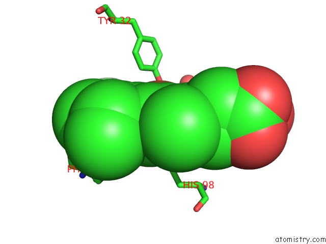
Mono view
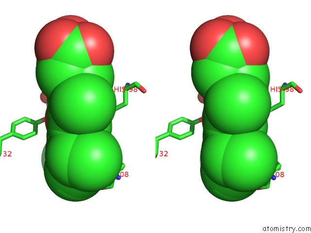
Stereo pair view
Mono view
Stereo pair view
A full contact list of Iron with other atoms in the Fe binding
site number 1 of Structural Plasticity in the Eight-Helix Fold of A Trematode Hemoglobin within 5.0Å range:
|
Reference:
M.Milani,
A.Pesce,
S.Dewilde,
P.Ascenzi,
L.Moens,
M.Bolognesi.
Structural Plasticity in the Eight-Helix Fold of A Trematode Haemoglobin. Acta Crystallogr.,Sect.D V. 58 719 2002.
ISSN: ISSN 0907-4449
PubMed: 11914507
DOI: 10.1107/S0907444902001865
Page generated: Wed Jul 16 16:58:19 2025
ISSN: ISSN 0907-4449
PubMed: 11914507
DOI: 10.1107/S0907444902001865
Last articles
Zn in 2UXCZn in 2UXA
Zn in 2UXB
Zn in 2USH
Zn in 2UVL
Zn in 2UUB
Zn in 2UUC
Zn in 2UUR
Zn in 2USN
Zn in 2UU9