Iron »
PDB 1k2r-1kqg »
1kok »
Iron in PDB 1kok: Crystal Structure of Mesopone Cytochrome C Peroxidase (Mpccp)
Enzymatic activity of Crystal Structure of Mesopone Cytochrome C Peroxidase (Mpccp)
All present enzymatic activity of Crystal Structure of Mesopone Cytochrome C Peroxidase (Mpccp):
1.11.1.5;
1.11.1.5;
Protein crystallography data
The structure of Crystal Structure of Mesopone Cytochrome C Peroxidase (Mpccp), PDB code: 1kok
was solved by
B.Bhaskar,
C.E.Immoos,
M.S.Cohen,
T.P.Barrows,
P.J.Farmer,
T.L.Poulos,
with X-Ray Crystallography technique. A brief refinement statistics is given in the table below:
| Resolution Low / High (Å) | 50.00 / 1.70 |
| Space group | P 21 21 21 |
| Cell size a, b, c (Å), α, β, γ (°) | 107.011, 76.091, 51.124, 90.00, 90.00, 90.00 |
| R / Rfree (%) | 18.6 / 20.7 |
Iron Binding Sites:
The binding sites of Iron atom in the Crystal Structure of Mesopone Cytochrome C Peroxidase (Mpccp)
(pdb code 1kok). This binding sites where shown within
5.0 Angstroms radius around Iron atom.
In total only one binding site of Iron was determined in the Crystal Structure of Mesopone Cytochrome C Peroxidase (Mpccp), PDB code: 1kok:
In total only one binding site of Iron was determined in the Crystal Structure of Mesopone Cytochrome C Peroxidase (Mpccp), PDB code: 1kok:
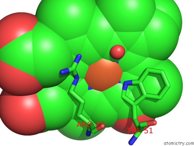
Iron binding site 1 out of 1 in 1kok
Go back to
Iron binding site 1 out
of 1 in the Crystal Structure of Mesopone Cytochrome C Peroxidase (Mpccp)
Mono view

Stereo pair view
Mono view
Stereo pair view
A full contact list of Iron with other atoms in the Fe binding
site number 1 of Crystal Structure of Mesopone Cytochrome C Peroxidase (Mpccp) within 5.0Å range:
|
Reference:
C.E.Immoos,
B.Bhaskar,
M.S.Cohen,
T.P.Barrows,
P.J.Farmer,
T.L.Poulos.
Mesopone Cytochrome C Peroxidase: Functional Model of Heme Oxygenated Oxidases. J.Inorg.Biochem. V. 91 635 2002.
ISSN: ISSN 0162-0134
PubMed: 12237229
DOI: 10.1016/S0162-0134(02)00447-6
Page generated: Wed Jul 16 17:07:44 2025
ISSN: ISSN 0162-0134
PubMed: 12237229
DOI: 10.1016/S0162-0134(02)00447-6
Last articles
Zn in 2R2LZn in 2R2H
Zn in 2R1W
Zn in 2R1X
Zn in 2R1Y
Zn in 2R23
Zn in 2QYV
Zn in 2QYN
Zn in 2QZR
Zn in 2QYK