Iron »
PDB 1kqj-1lfg »
1kqj »
Iron in PDB 1kqj: Crystal Structure of A Mutant of Muty Catalytic Domain
Protein crystallography data
The structure of Crystal Structure of A Mutant of Muty Catalytic Domain, PDB code: 1kqj
was solved by
T.E.Messick,
N.H.Chmiel,
M.P.Golinelli,
S.S.David,
L.Joshua-Tor,
with X-Ray Crystallography technique. A brief refinement statistics is given in the table below:
| Resolution Low / High (Å) | 29.65 / 1.70 |
| Space group | C 1 2 1 |
| Cell size a, b, c (Å), α, β, γ (°) | 83.550, 49.900, 71.010, 90.00, 122.56, 90.00 |
| R / Rfree (%) | 19.1 / 20.8 |
Iron Binding Sites:
The binding sites of Iron atom in the Crystal Structure of A Mutant of Muty Catalytic Domain
(pdb code 1kqj). This binding sites where shown within
5.0 Angstroms radius around Iron atom.
In total 4 binding sites of Iron where determined in the Crystal Structure of A Mutant of Muty Catalytic Domain, PDB code: 1kqj:
Jump to Iron binding site number: 1; 2; 3; 4;
In total 4 binding sites of Iron where determined in the Crystal Structure of A Mutant of Muty Catalytic Domain, PDB code: 1kqj:
Jump to Iron binding site number: 1; 2; 3; 4;
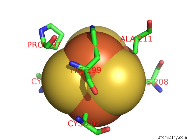
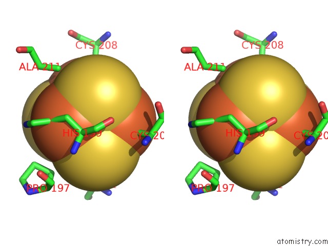
Iron binding site 1 out of 4 in 1kqj
Go back to
Iron binding site 1 out
of 4 in the Crystal Structure of A Mutant of Muty Catalytic Domain
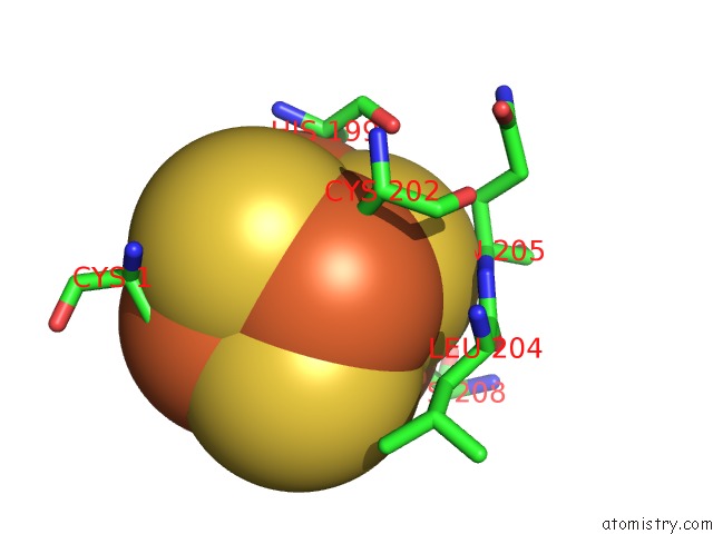
Mono view
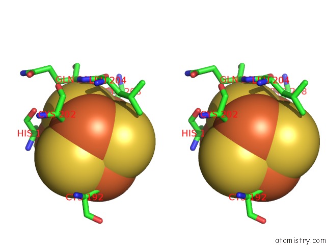
Stereo pair view
Mono view
Stereo pair view
A full contact list of Iron with other atoms in the Fe binding
site number 1 of Crystal Structure of A Mutant of Muty Catalytic Domain within 5.0Å range:
|
Iron binding site 2 out of 4 in 1kqj
Go back to
Iron binding site 2 out
of 4 in the Crystal Structure of A Mutant of Muty Catalytic Domain
Mono view
Stereo pair view
Mono view
Stereo pair view
A full contact list of Iron with other atoms in the Fe binding
site number 2 of Crystal Structure of A Mutant of Muty Catalytic Domain within 5.0Å range:
|
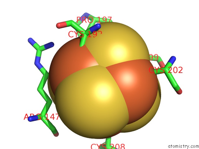
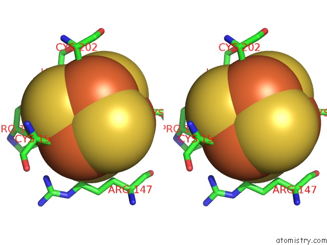
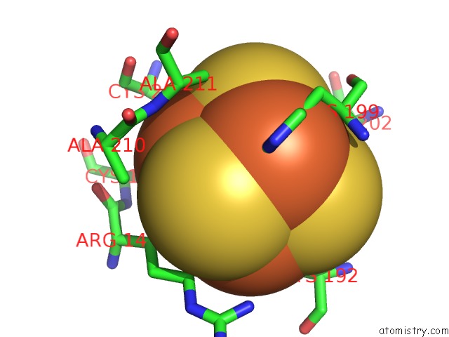
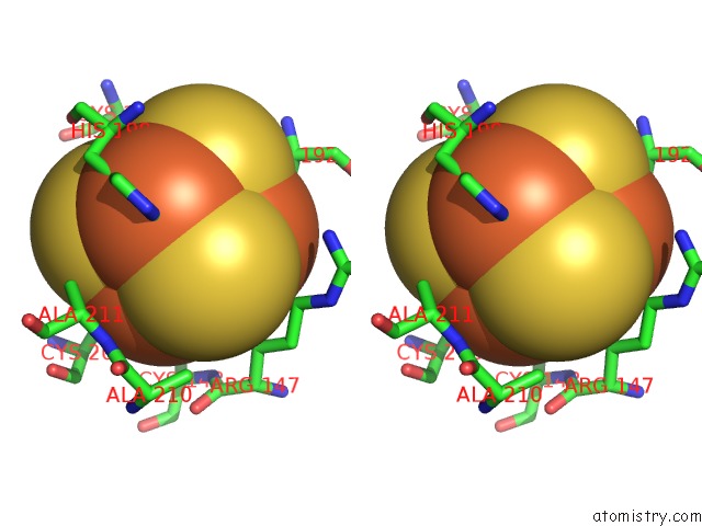
Iron binding site 3 out of 4 in 1kqj
Go back to
Iron binding site 3 out
of 4 in the Crystal Structure of A Mutant of Muty Catalytic Domain
Mono view
Stereo pair view
Mono view
Stereo pair view
A full contact list of Iron with other atoms in the Fe binding
site number 3 of Crystal Structure of A Mutant of Muty Catalytic Domain within 5.0Å range:
|
Iron binding site 4 out of 4 in 1kqj
Go back to
Iron binding site 4 out
of 4 in the Crystal Structure of A Mutant of Muty Catalytic Domain
Mono view
Stereo pair view
Mono view
Stereo pair view
A full contact list of Iron with other atoms in the Fe binding
site number 4 of Crystal Structure of A Mutant of Muty Catalytic Domain within 5.0Å range:
|
Reference:
T.E.Messick,
N.H.Chmiel,
M.P.Golinelli,
M.R.Langer,
L.Joshua-Tor,
S.S.David.
Noncysteinyl Coordination to the [4FE-4S]2+ Cluster of the Dna Repair Adenine Glycosylase Muty Introduced Via Site-Directed Mutagenesis. Structural Characterization of An Unusual Histidinyl-Coordinated Cluster. Biochemistry V. 41 3931 2002.
ISSN: ISSN 0006-2960
PubMed: 11900536
DOI: 10.1021/BI012035X
Page generated: Wed Jul 16 17:11:58 2025
ISSN: ISSN 0006-2960
PubMed: 11900536
DOI: 10.1021/BI012035X
Last articles
W in 9FPPW in 8PRM
W in 9QM1
W in 9QM0
W in 9OJ3
W in 9MQX
W in 9FP4
W in 9BEO
W in 9BEM
W in 8P2U