Iron »
PDB 1kqj-1lfg »
1lco »
Iron in PDB 1lco: X-Ray Structure of Two Complexes of the Y143F Flavocytochrome B2 Mutant Crystallized in the Presence of Lactate or Phenyl-Lactate
Enzymatic activity of X-Ray Structure of Two Complexes of the Y143F Flavocytochrome B2 Mutant Crystallized in the Presence of Lactate or Phenyl-Lactate
All present enzymatic activity of X-Ray Structure of Two Complexes of the Y143F Flavocytochrome B2 Mutant Crystallized in the Presence of Lactate or Phenyl-Lactate:
1.1.2.3;
1.1.2.3;
Protein crystallography data
The structure of X-Ray Structure of Two Complexes of the Y143F Flavocytochrome B2 Mutant Crystallized in the Presence of Lactate or Phenyl-Lactate, PDB code: 1lco
was solved by
M.Tegoni,
C.Cambillau,
with X-Ray Crystallography technique. A brief refinement statistics is given in the table below:
| Resolution Low / High (Å) | 6.00 / 2.90 |
| Space group | P 32 2 1 |
| Cell size a, b, c (Å), α, β, γ (°) | 164.500, 164.500, 114.000, 90.00, 90.00, 120.00 |
| R / Rfree (%) | 18.6 / n/a |
Iron Binding Sites:
The binding sites of Iron atom in the X-Ray Structure of Two Complexes of the Y143F Flavocytochrome B2 Mutant Crystallized in the Presence of Lactate or Phenyl-Lactate
(pdb code 1lco). This binding sites where shown within
5.0 Angstroms radius around Iron atom.
In total only one binding site of Iron was determined in the X-Ray Structure of Two Complexes of the Y143F Flavocytochrome B2 Mutant Crystallized in the Presence of Lactate or Phenyl-Lactate, PDB code: 1lco:
In total only one binding site of Iron was determined in the X-Ray Structure of Two Complexes of the Y143F Flavocytochrome B2 Mutant Crystallized in the Presence of Lactate or Phenyl-Lactate, PDB code: 1lco:
Iron binding site 1 out of 1 in 1lco
Go back to
Iron binding site 1 out
of 1 in the X-Ray Structure of Two Complexes of the Y143F Flavocytochrome B2 Mutant Crystallized in the Presence of Lactate or Phenyl-Lactate
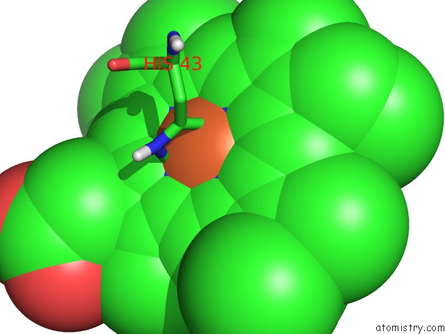
Mono view
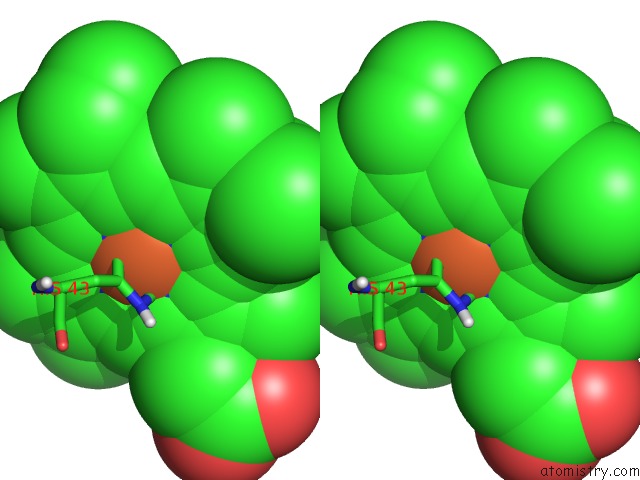
Stereo pair view
Mono view
Stereo pair view
A full contact list of Iron with other atoms in the Fe binding
site number 1 of X-Ray Structure of Two Complexes of the Y143F Flavocytochrome B2 Mutant Crystallized in the Presence of Lactate or Phenyl-Lactate within 5.0Å range:
|
Reference:
M.Tegoni,
S.Begotti,
C.Cambillau.
X-Ray Structure of Two Complexes of the Y143F Flavocytochrome B2 Mutant Crystallized in the Presence of Lactate or Phenyl Lactate. Biochemistry V. 34 9840 1995.
ISSN: ISSN 0006-2960
PubMed: 7632684
DOI: 10.1021/BI00031A004
Page generated: Wed Jul 16 17:22:17 2025
ISSN: ISSN 0006-2960
PubMed: 7632684
DOI: 10.1021/BI00031A004
Last articles
Zn in 1SG0Zn in 1SG6
Zn in 1SED
Zn in 1SE0
Zn in 1SDZ
Zn in 1S3Q
Zn in 1SDY
Zn in 1S7G
Zn in 1SDA
Zn in 1SDX