Iron »
PDB 1lrm-1m6m »
1ltv »
Iron in PDB 1ltv: Crystal Structure of Chromobacterium Violaceum Phenylalanine Hydroxylase, Structure with Bound Oxidized Fe(III)
Enzymatic activity of Crystal Structure of Chromobacterium Violaceum Phenylalanine Hydroxylase, Structure with Bound Oxidized Fe(III)
All present enzymatic activity of Crystal Structure of Chromobacterium Violaceum Phenylalanine Hydroxylase, Structure with Bound Oxidized Fe(III):
1.14.16.1;
1.14.16.1;
Protein crystallography data
The structure of Crystal Structure of Chromobacterium Violaceum Phenylalanine Hydroxylase, Structure with Bound Oxidized Fe(III), PDB code: 1ltv
was solved by
H.Erlandsen,
J.Y.Kim,
M.G.Patch,
A.Han,
A.Volner,
M.M.Abu-Omar,
R.C.Stevens,
with X-Ray Crystallography technique. A brief refinement statistics is given in the table below:
| Resolution Low / High (Å) | 20.00 / 2.00 |
| Space group | P 21 21 21 |
| Cell size a, b, c (Å), α, β, γ (°) | 46.594, 67.533, 90.847, 90.00, 90.00, 90.00 |
| R / Rfree (%) | 20.7 / 26.1 |
Iron Binding Sites:
The binding sites of Iron atom in the Crystal Structure of Chromobacterium Violaceum Phenylalanine Hydroxylase, Structure with Bound Oxidized Fe(III)
(pdb code 1ltv). This binding sites where shown within
5.0 Angstroms radius around Iron atom.
In total only one binding site of Iron was determined in the Crystal Structure of Chromobacterium Violaceum Phenylalanine Hydroxylase, Structure with Bound Oxidized Fe(III), PDB code: 1ltv:
In total only one binding site of Iron was determined in the Crystal Structure of Chromobacterium Violaceum Phenylalanine Hydroxylase, Structure with Bound Oxidized Fe(III), PDB code: 1ltv:
Iron binding site 1 out of 1 in 1ltv
Go back to
Iron binding site 1 out
of 1 in the Crystal Structure of Chromobacterium Violaceum Phenylalanine Hydroxylase, Structure with Bound Oxidized Fe(III)
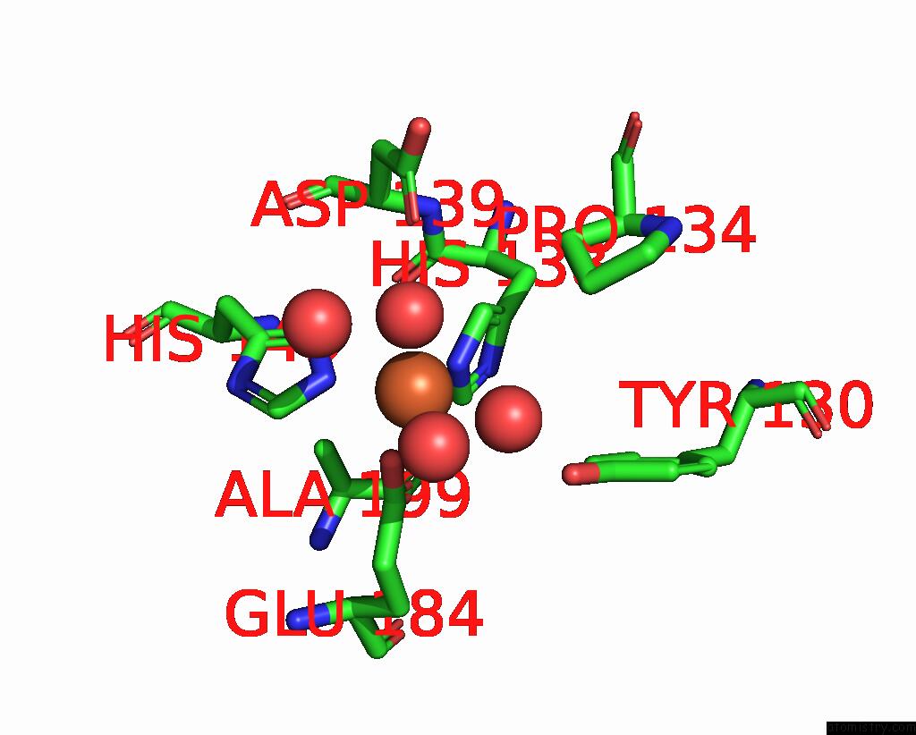
Mono view
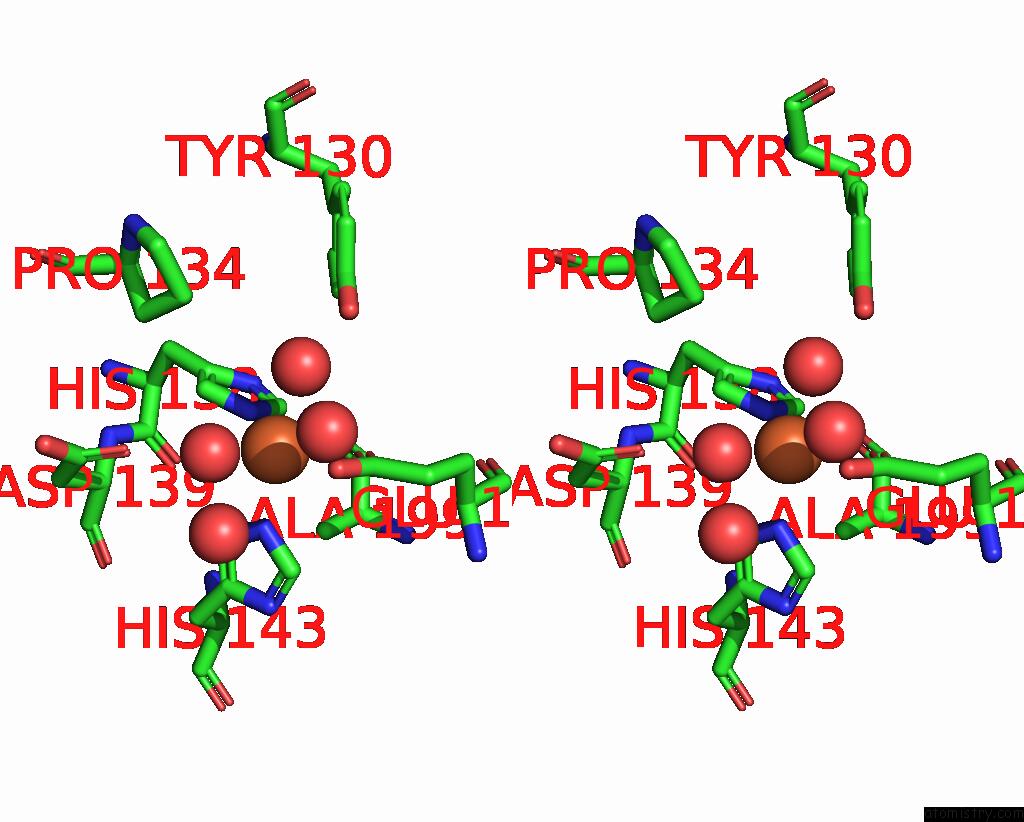
Stereo pair view
Mono view
Stereo pair view
A full contact list of Iron with other atoms in the Fe binding
site number 1 of Crystal Structure of Chromobacterium Violaceum Phenylalanine Hydroxylase, Structure with Bound Oxidized Fe(III) within 5.0Å range:
|
Reference:
H.Erlandsen,
J.Y.Kim,
M.G.Patch,
A.Han,
A.Volner,
M.M.Abu-Omar,
R.C.Stevens.
Structural Comparison of Bacterial and Human Iron-Dependent Phenylalanine Hydroxylases: Similar Fold, Different Stability and Reaction Rates. J.Mol.Biol. V. 320 645 2002.
ISSN: ISSN 0022-2836
PubMed: 12096915
DOI: 10.1016/S0022-2836(02)00496-5
Page generated: Wed Jul 16 17:29:52 2025
ISSN: ISSN 0022-2836
PubMed: 12096915
DOI: 10.1016/S0022-2836(02)00496-5
Last articles
Mg in 4DR7Mg in 4DR6
Mg in 4DR5
Mg in 4DUX
Mg in 4DUW
Mg in 4DUV
Mg in 4DUO
Mg in 4DUG
Mg in 4DTY
Mg in 4DTW