Iron »
PDB 1lrm-1m6m »
1m20 »
Iron in PDB 1m20: Crystal Structure of F35Y Mutant of Trypsin-Solubilized Fragment of Cytochrome B5
Protein crystallography data
The structure of Crystal Structure of F35Y Mutant of Trypsin-Solubilized Fragment of Cytochrome B5, PDB code: 1m20
was solved by
P.Yao,
J.Wu,
Y.-H.Wang,
B.-Y.Sun,
Z.-X.Xia,
Z.-X.Huang,
with X-Ray Crystallography technique. A brief refinement statistics is given in the table below:
| Resolution Low / High (Å) | 19.91 / 1.80 |
| Space group | C 1 2 1 |
| Cell size a, b, c (Å), α, β, γ (°) | 70.709, 40.388, 39.299, 90.00, 111.72, 90.00 |
| R / Rfree (%) | 19.3 / 22.7 |
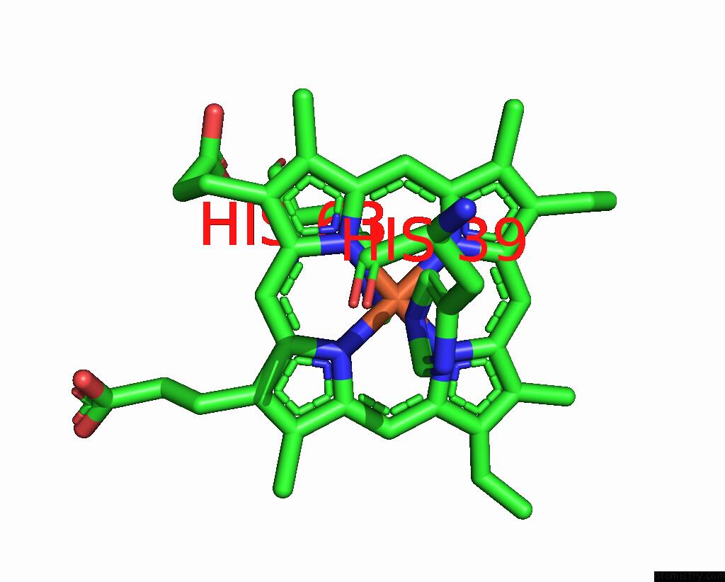
Iron Binding Sites:
The binding sites of Iron atom in the Crystal Structure of F35Y Mutant of Trypsin-Solubilized Fragment of Cytochrome B5
(pdb code 1m20). This binding sites where shown within
5.0 Angstroms radius around Iron atom.
In total only one binding site of Iron was determined in the Crystal Structure of F35Y Mutant of Trypsin-Solubilized Fragment of Cytochrome B5, PDB code: 1m20:
In total only one binding site of Iron was determined in the Crystal Structure of F35Y Mutant of Trypsin-Solubilized Fragment of Cytochrome B5, PDB code: 1m20:
Iron binding site 1 out of 1 in 1m20
Go back to
Iron binding site 1 out
of 1 in the Crystal Structure of F35Y Mutant of Trypsin-Solubilized Fragment of Cytochrome B5
Mono view

Stereo pair view
Mono view
Stereo pair view
A full contact list of Iron with other atoms in the Fe binding
site number 1 of Crystal Structure of F35Y Mutant of Trypsin-Solubilized Fragment of Cytochrome B5 within 5.0Å range:
|
Reference:
P.Yao,
J.Wu,
Y.H.Wang,
B.Y.Sun,
Z.X.Xia,
Z.X.Huang.
X-Ray Crystallography, Cd and Kinetic Studies Revealed the Essence of the Abnormal Behaviors of the Cytochrome B5 PHE35-->Tyr Mutant. Eur.J.Biochem. V. 269 4287 2002.
ISSN: ISSN 0014-2956
PubMed: 12199707
DOI: 10.1046/J.1432-1033.2002.03120.X
Page generated: Wed Jul 16 17:33:01 2025
ISSN: ISSN 0014-2956
PubMed: 12199707
DOI: 10.1046/J.1432-1033.2002.03120.X
Last articles
Mg in 4B48Mg in 4B47
Mg in 4B43
Mg in 4B3S
Mg in 4B2Q
Mg in 4B3A
Mg in 4B1Z
Mg in 4B2P
Mg in 4B2M
Mg in 4B2K