Iron »
PDB 1m6z-1mko »
1mbc »
Iron in PDB 1mbc: X-Ray Structure and Refinement of Carbon-Monoxy (Fe II)- Myoglobin at 1.5 Angstroms Resolution
Protein crystallography data
The structure of X-Ray Structure and Refinement of Carbon-Monoxy (Fe II)- Myoglobin at 1.5 Angstroms Resolution, PDB code: 1mbc
was solved by
J.Kuriyan,
G.A.Petsko,
with X-Ray Crystallography technique. A brief refinement statistics is given in the table below:
| Resolution Low / High (Å) | N/A / 1.50 |
| Space group | P 1 21 1 |
| Cell size a, b, c (Å), α, β, γ (°) | 64.180, 30.840, 34.690, 90.00, 105.84, 90.00 |
| R / Rfree (%) | n/a / n/a |
Iron Binding Sites:
The binding sites of Iron atom in the X-Ray Structure and Refinement of Carbon-Monoxy (Fe II)- Myoglobin at 1.5 Angstroms Resolution
(pdb code 1mbc). This binding sites where shown within
5.0 Angstroms radius around Iron atom.
In total only one binding site of Iron was determined in the X-Ray Structure and Refinement of Carbon-Monoxy (Fe II)- Myoglobin at 1.5 Angstroms Resolution, PDB code: 1mbc:
In total only one binding site of Iron was determined in the X-Ray Structure and Refinement of Carbon-Monoxy (Fe II)- Myoglobin at 1.5 Angstroms Resolution, PDB code: 1mbc:
Iron binding site 1 out of 1 in 1mbc
Go back to
Iron binding site 1 out
of 1 in the X-Ray Structure and Refinement of Carbon-Monoxy (Fe II)- Myoglobin at 1.5 Angstroms Resolution
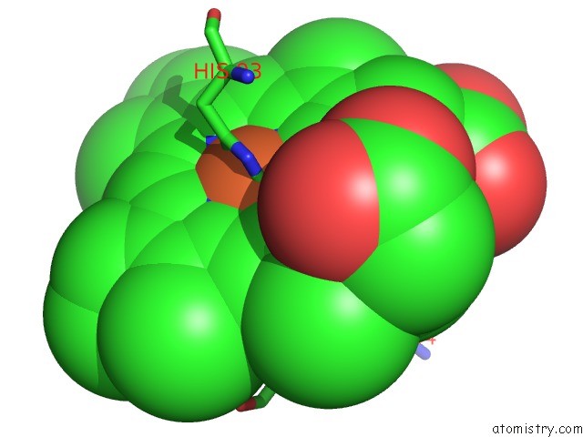
Mono view
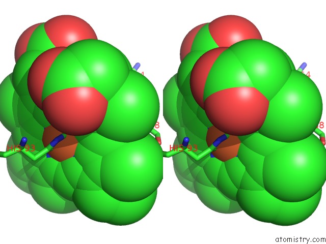
Stereo pair view
Mono view
Stereo pair view
A full contact list of Iron with other atoms in the Fe binding
site number 1 of X-Ray Structure and Refinement of Carbon-Monoxy (Fe II)- Myoglobin at 1.5 Angstroms Resolution within 5.0Å range:
|
Reference:
J.Kuriyan,
S.Wilz,
M.Karplus,
G.A.Petsko.
X-Ray Structure and Refinement of Carbon-Monoxy (Fe II)-Myoglobin at 1.5 A Resolution. J.Mol.Biol. V. 192 133 1986.
ISSN: ISSN 0022-2836
PubMed: 3820301
DOI: 10.1016/0022-2836(86)90470-5
Page generated: Wed Jul 16 18:01:20 2025
ISSN: ISSN 0022-2836
PubMed: 3820301
DOI: 10.1016/0022-2836(86)90470-5
Last articles
K in 1JFVK in 1JF8
K in 1JDR
K in 1JCI
K in 1JBS
K in 1JBT
K in 1J95
K in 1JBR
K in 1J5Y
K in 1IS8