Iron »
PDB 1m6z-1mko »
1mhz »
Iron in PDB 1mhz: Methane Monooxygenase Hydroxylase
Enzymatic activity of Methane Monooxygenase Hydroxylase
All present enzymatic activity of Methane Monooxygenase Hydroxylase:
1.14.13.25;
1.14.13.25;
Protein crystallography data
The structure of Methane Monooxygenase Hydroxylase, PDB code: 1mhz
was solved by
N.Elango,
R.Radhakrishnan,
W.A.Froland,
B.J.Waller,
C.A.Earhart,
J.D.Lipscomb,
D.H.Ohlendorf,
with X-Ray Crystallography technique. A brief refinement statistics is given in the table below:
| Resolution Low / High (Å) | 5.00 / 2.70 |
| Space group | C 2 2 21 |
| Cell size a, b, c (Å), α, β, γ (°) | 293.380, 64.010, 143.650, 90.00, 90.00, 90.00 |
| R / Rfree (%) | 15.2 / n/a |
Iron Binding Sites:
The binding sites of Iron atom in the Methane Monooxygenase Hydroxylase
(pdb code 1mhz). This binding sites where shown within
5.0 Angstroms radius around Iron atom.
In total 2 binding sites of Iron where determined in the Methane Monooxygenase Hydroxylase, PDB code: 1mhz:
Jump to Iron binding site number: 1; 2;
In total 2 binding sites of Iron where determined in the Methane Monooxygenase Hydroxylase, PDB code: 1mhz:
Jump to Iron binding site number: 1; 2;
Iron binding site 1 out of 2 in 1mhz
Go back to
Iron binding site 1 out
of 2 in the Methane Monooxygenase Hydroxylase

Mono view
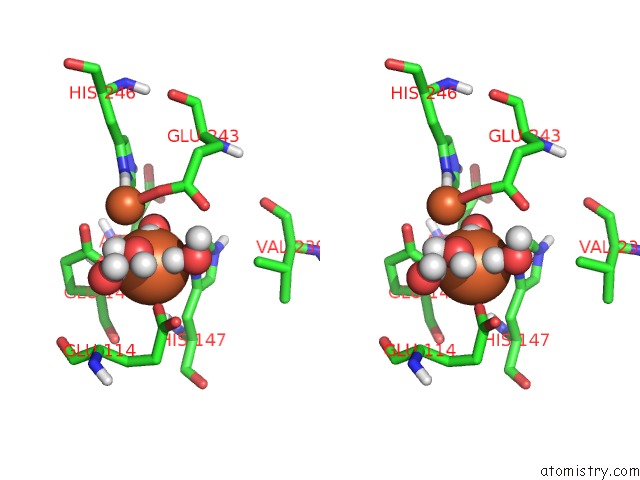
Stereo pair view
Mono view
Stereo pair view
A full contact list of Iron with other atoms in the Fe binding
site number 1 of Methane Monooxygenase Hydroxylase within 5.0Å range:
|
Iron binding site 2 out of 2 in 1mhz
Go back to
Iron binding site 2 out
of 2 in the Methane Monooxygenase Hydroxylase
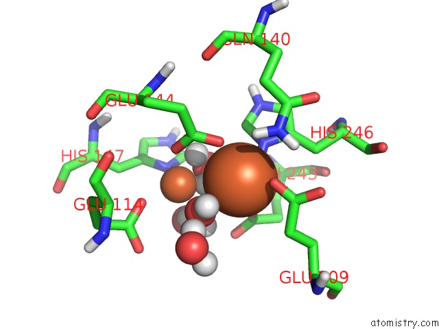
Mono view

Stereo pair view
Mono view
Stereo pair view
A full contact list of Iron with other atoms in the Fe binding
site number 2 of Methane Monooxygenase Hydroxylase within 5.0Å range:
|
Reference:
N.Elango,
R.Radhakrishnan,
W.A.Froland,
B.J.Wallar,
C.A.Earhart,
J.D.Lipscomb,
D.H.Ohlendorf.
Crystal Structure of the Hydroxylase Component of Methane Monooxygenase From Methylosinus Trichosporium OB3B Protein Sci. V. 6 556 1997.
ISSN: ISSN 0961-8368
PubMed: 9070438
Page generated: Wed Jul 16 18:03:40 2025
ISSN: ISSN 0961-8368
PubMed: 9070438
Last articles
K in 6SE7K in 6SE5
K in 6SBI
K in 6RVZ
K in 6S15
K in 6RW0
K in 6RWV
K in 6RV4
K in 6RV3
K in 6RV2