Iron »
PDB 1mkq-1mpw »
1mkr »
Iron in PDB 1mkr: Crystal Structure of A Mutant Variant of Cytochrome C Peroxidase (Plate Like Crystals)
Enzymatic activity of Crystal Structure of A Mutant Variant of Cytochrome C Peroxidase (Plate Like Crystals)
All present enzymatic activity of Crystal Structure of A Mutant Variant of Cytochrome C Peroxidase (Plate Like Crystals):
1.11.1.5;
1.11.1.5;
Protein crystallography data
The structure of Crystal Structure of A Mutant Variant of Cytochrome C Peroxidase (Plate Like Crystals), PDB code: 1mkr
was solved by
B.Bhaskar,
C.E.Immoos,
H.Shimizu,
P.J.Farmer,
T.L.Poulos,
with X-Ray Crystallography technique. A brief refinement statistics is given in the table below:
| Resolution Low / High (Å) | 50.00 / 1.58 |
| Space group | P 21 21 21 |
| Cell size a, b, c (Å), α, β, γ (°) | 50.480, 50.957, 118.884, 90.00, 90.00, 90.00 |
| R / Rfree (%) | 19.4 / 21.9 |
Iron Binding Sites:
The binding sites of Iron atom in the Crystal Structure of A Mutant Variant of Cytochrome C Peroxidase (Plate Like Crystals)
(pdb code 1mkr). This binding sites where shown within
5.0 Angstroms radius around Iron atom.
In total only one binding site of Iron was determined in the Crystal Structure of A Mutant Variant of Cytochrome C Peroxidase (Plate Like Crystals), PDB code: 1mkr:
In total only one binding site of Iron was determined in the Crystal Structure of A Mutant Variant of Cytochrome C Peroxidase (Plate Like Crystals), PDB code: 1mkr:
Iron binding site 1 out of 1 in 1mkr
Go back to
Iron binding site 1 out
of 1 in the Crystal Structure of A Mutant Variant of Cytochrome C Peroxidase (Plate Like Crystals)
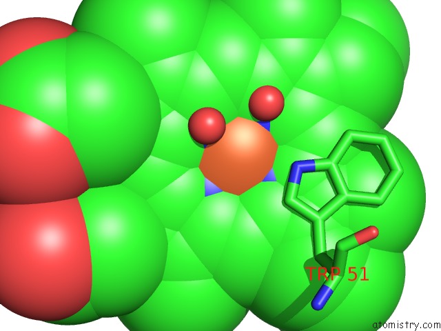
Mono view
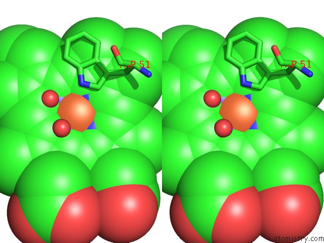
Stereo pair view
Mono view
Stereo pair view
A full contact list of Iron with other atoms in the Fe binding
site number 1 of Crystal Structure of A Mutant Variant of Cytochrome C Peroxidase (Plate Like Crystals) within 5.0Å range:
|
Reference:
B.Bhaskar,
C.E.Immoos,
H.Shimizu,
F.Sulc,
P.J.Farmer,
T.L.Poulos.
A Novel Heme and Peroxide-Dependent Tryptophan-Tyrosine Cross-Link in A Mutant of Cytochrome C Peroxidase J.Mol.Biol. V. 328 157 2003.
ISSN: ISSN 0022-2836
PubMed: 12684005
DOI: 10.1016/S0022-2836(03)00179-7
Page generated: Wed Jul 16 18:13:19 2025
ISSN: ISSN 0022-2836
PubMed: 12684005
DOI: 10.1016/S0022-2836(03)00179-7
Last articles
Mg in 5XYUMg in 5Y4N
Mg in 5Y4J
Mg in 5Y4I
Mg in 5Y3J
Mg in 5Y41
Mg in 5Y3I
Mg in 5Y36
Mg in 5Y34
Mg in 5Y1W