Iron »
PDB 1mkq-1mpw »
1mn1 »
Iron in PDB 1mn1: Manganese Peroxidase Substrate Binding Site Mutant D179N
Enzymatic activity of Manganese Peroxidase Substrate Binding Site Mutant D179N
All present enzymatic activity of Manganese Peroxidase Substrate Binding Site Mutant D179N:
1.11.1.13;
1.11.1.13;
Protein crystallography data
The structure of Manganese Peroxidase Substrate Binding Site Mutant D179N, PDB code: 1mn1
was solved by
M.Sundaramoorthy,
T.L.Poulos,
with X-Ray Crystallography technique. A brief refinement statistics is given in the table below:
| Resolution Low / High (Å) | 8.00 / 2.00 |
| Space group | C 1 2 1 |
| Cell size a, b, c (Å), α, β, γ (°) | 163.240, 45.970, 53.570, 90.00, 97.16, 90.00 |
| R / Rfree (%) | 18.7 / n/a |
Other elements in 1mn1:
The structure of Manganese Peroxidase Substrate Binding Site Mutant D179N also contains other interesting chemical elements:
| Calcium | (Ca) | 2 atoms |
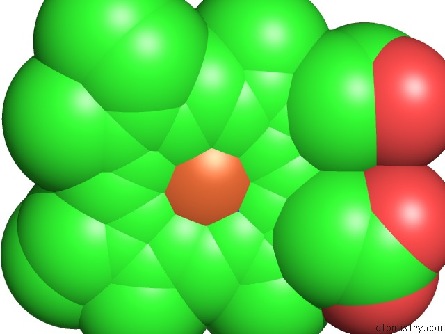
Iron Binding Sites:
The binding sites of Iron atom in the Manganese Peroxidase Substrate Binding Site Mutant D179N
(pdb code 1mn1). This binding sites where shown within
5.0 Angstroms radius around Iron atom.
In total only one binding site of Iron was determined in the Manganese Peroxidase Substrate Binding Site Mutant D179N, PDB code: 1mn1:
In total only one binding site of Iron was determined in the Manganese Peroxidase Substrate Binding Site Mutant D179N, PDB code: 1mn1:
Iron binding site 1 out of 1 in 1mn1
Go back to
Iron binding site 1 out
of 1 in the Manganese Peroxidase Substrate Binding Site Mutant D179N
Mono view

Stereo pair view
Mono view
Stereo pair view
A full contact list of Iron with other atoms in the Fe binding
site number 1 of Manganese Peroxidase Substrate Binding Site Mutant D179N within 5.0Å range:
|
Reference:
M.Sundaramoorthy,
K.Kishi,
M.H.Gold,
T.L.Poulos.
Crystal Structures of Substrate Binding Site Mutants of Manganese Peroxidase. J.Biol.Chem. V. 272 17574 1997.
ISSN: ISSN 0021-9258
PubMed: 9211904
DOI: 10.1074/JBC.272.28.17574
Page generated: Wed Jul 16 18:15:09 2025
ISSN: ISSN 0021-9258
PubMed: 9211904
DOI: 10.1074/JBC.272.28.17574
Last articles
Mg in 4WFNMg in 4WF9
Mg in 4WFB
Mg in 4WFM
Mg in 4WFL
Mg in 4WCE
Mg in 4WF7
Mg in 4WEC
Mg in 4WEO
Mg in 4WCW