Iron »
PDB 1mkq-1mpw »
1moh »
Iron in PDB 1moh: Ferric Monomeric Hemoglobin I (Hb I)
Protein crystallography data
The structure of Ferric Monomeric Hemoglobin I (Hb I), PDB code: 1moh
was solved by
M.Rizzi,
J.B.Wittenberg,
P.Ascenzi,
M.Bolognesi,
with X-Ray Crystallography technique. A brief refinement statistics is given in the table below:
| Resolution Low / High (Å) | 15.00 / 1.90 |
| Space group | P 1 21 1 |
| Cell size a, b, c (Å), α, β, γ (°) | 50.550, 38.790, 42.510, 90.00, 106.93, 90.00 |
| R / Rfree (%) | 18.6 / n/a |
Iron Binding Sites:
The binding sites of Iron atom in the Ferric Monomeric Hemoglobin I (Hb I)
(pdb code 1moh). This binding sites where shown within
5.0 Angstroms radius around Iron atom.
In total only one binding site of Iron was determined in the Ferric Monomeric Hemoglobin I (Hb I), PDB code: 1moh:
In total only one binding site of Iron was determined in the Ferric Monomeric Hemoglobin I (Hb I), PDB code: 1moh:
Iron binding site 1 out of 1 in 1moh
Go back to
Iron binding site 1 out
of 1 in the Ferric Monomeric Hemoglobin I (Hb I)
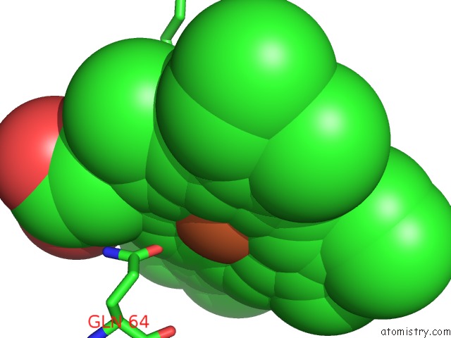
Mono view
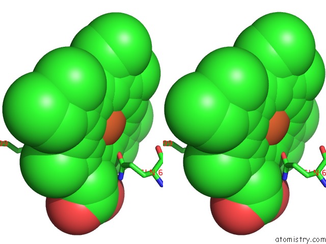
Stereo pair view
Mono view
Stereo pair view
A full contact list of Iron with other atoms in the Fe binding
site number 1 of Ferric Monomeric Hemoglobin I (Hb I) within 5.0Å range:
|
Reference:
M.Rizzi,
J.B.Wittenberg,
A.Coda,
P.Ascenzi,
M.Bolognesi.
Structural Bases For Sulfide Recognition in Lucina Pectinata Hemoglobin I. J.Mol.Biol. V. 258 1 1996.
ISSN: ISSN 0022-2836
PubMed: 8613980
DOI: 10.1006/JMBI.1996.0228
Page generated: Wed Jul 16 18:16:55 2025
ISSN: ISSN 0022-2836
PubMed: 8613980
DOI: 10.1006/JMBI.1996.0228
Last articles
Mg in 6CA4Mg in 6C90
Mg in 6CA0
Mg in 6C9Y
Mg in 6C8Z
Mg in 6C8P
Mg in 6C8N
Mg in 6C8O
Mg in 6C8D
Mg in 6C8L