Iron »
PDB 1n5w-1nmi »
1n5w »
Iron in PDB 1n5w: Crystal Structure of the Cu,Mo-Co Dehydrogenase (Codh); Oxidized Form
Enzymatic activity of Crystal Structure of the Cu,Mo-Co Dehydrogenase (Codh); Oxidized Form
All present enzymatic activity of Crystal Structure of the Cu,Mo-Co Dehydrogenase (Codh); Oxidized Form:
1.2.99.2;
1.2.99.2;
Protein crystallography data
The structure of Crystal Structure of the Cu,Mo-Co Dehydrogenase (Codh); Oxidized Form, PDB code: 1n5w
was solved by
H.Dobbek,
L.Gremer,
R.Kiefersauer,
R.Huber,
O.Meyer,
with X-Ray Crystallography technique. A brief refinement statistics is given in the table below:
| Resolution Low / High (Å) | 18.00 / 1.50 |
| Space group | P 21 21 21 |
| Cell size a, b, c (Å), α, β, γ (°) | 119.295, 132.088, 159.825, 90.00, 90.00, 90.00 |
| R / Rfree (%) | 13.5 / 17.1 |
Other elements in 1n5w:
The structure of Crystal Structure of the Cu,Mo-Co Dehydrogenase (Codh); Oxidized Form also contains other interesting chemical elements:
| Molybdenum | (Mo) | 2 atoms |
| Copper | (Cu) | 2 atoms |
Iron Binding Sites:
The binding sites of Iron atom in the Crystal Structure of the Cu,Mo-Co Dehydrogenase (Codh); Oxidized Form
(pdb code 1n5w). This binding sites where shown within
5.0 Angstroms radius around Iron atom.
In total 8 binding sites of Iron where determined in the Crystal Structure of the Cu,Mo-Co Dehydrogenase (Codh); Oxidized Form, PDB code: 1n5w:
Jump to Iron binding site number: 1; 2; 3; 4; 5; 6; 7; 8;
In total 8 binding sites of Iron where determined in the Crystal Structure of the Cu,Mo-Co Dehydrogenase (Codh); Oxidized Form, PDB code: 1n5w:
Jump to Iron binding site number: 1; 2; 3; 4; 5; 6; 7; 8;
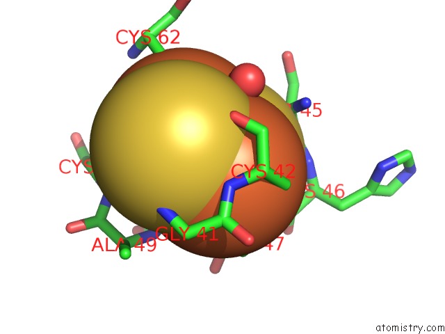
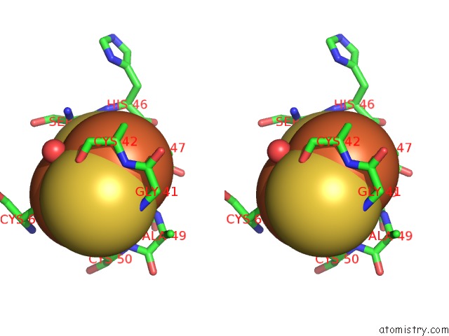
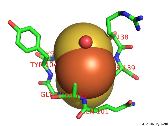
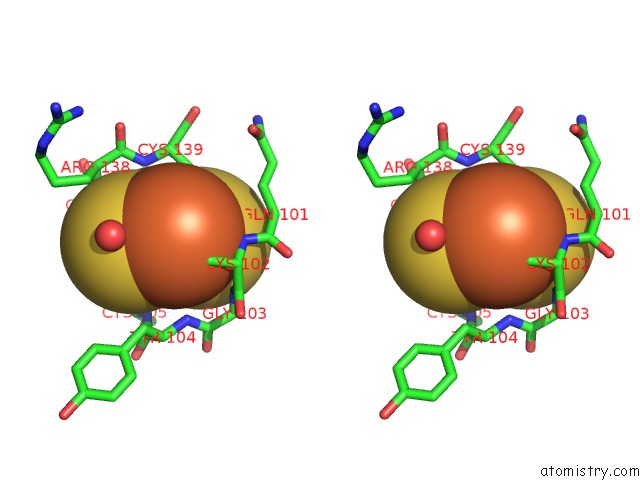
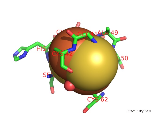
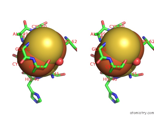
Iron binding site 1 out of 8 in 1n5w
Go back to
Iron binding site 1 out
of 8 in the Crystal Structure of the Cu,Mo-Co Dehydrogenase (Codh); Oxidized Form
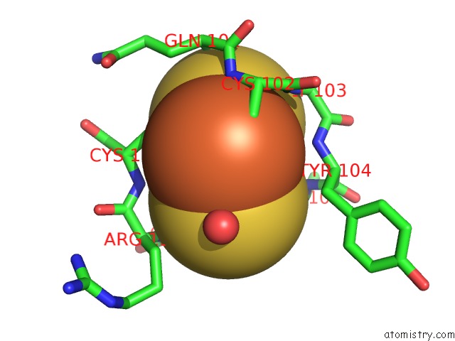
Mono view
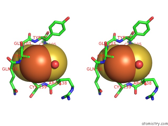
Stereo pair view
Mono view
Stereo pair view
A full contact list of Iron with other atoms in the Fe binding
site number 1 of Crystal Structure of the Cu,Mo-Co Dehydrogenase (Codh); Oxidized Form within 5.0Å range:
|
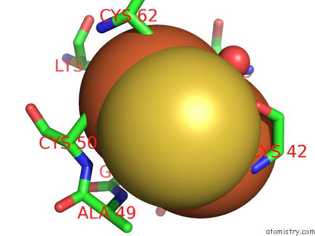
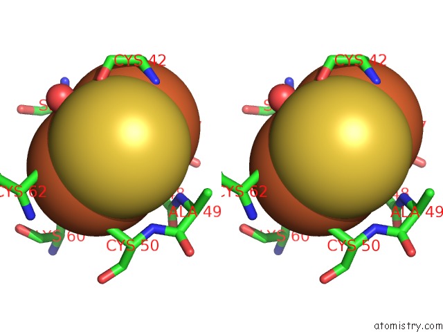
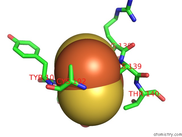
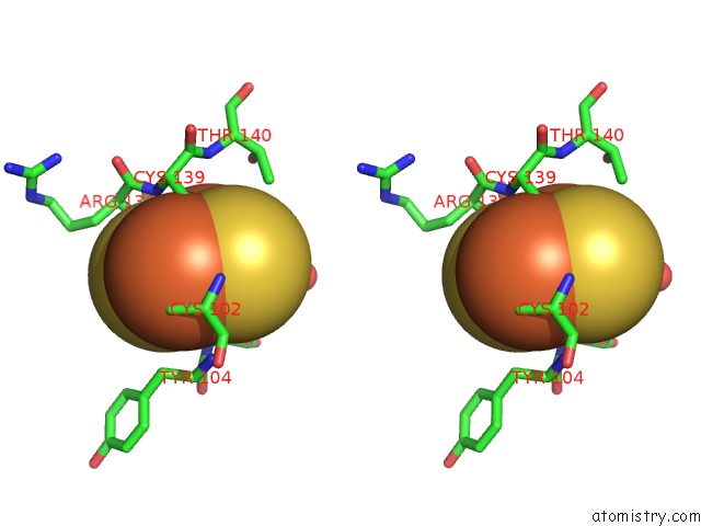
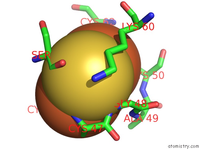
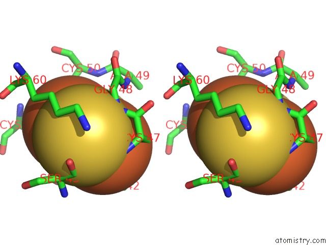
Iron binding site 2 out of 8 in 1n5w
Go back to
Iron binding site 2 out
of 8 in the Crystal Structure of the Cu,Mo-Co Dehydrogenase (Codh); Oxidized Form
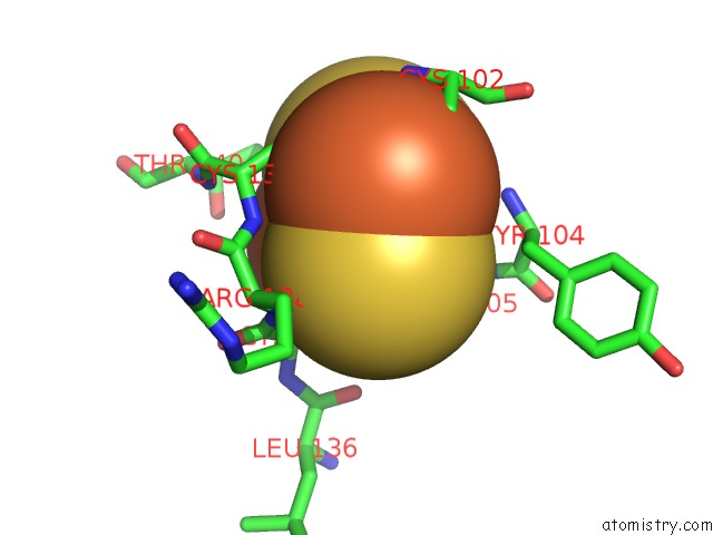
Mono view
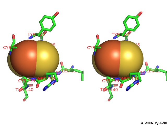
Stereo pair view
Mono view
Stereo pair view
A full contact list of Iron with other atoms in the Fe binding
site number 2 of Crystal Structure of the Cu,Mo-Co Dehydrogenase (Codh); Oxidized Form within 5.0Å range:
|
Iron binding site 3 out of 8 in 1n5w
Go back to
Iron binding site 3 out
of 8 in the Crystal Structure of the Cu,Mo-Co Dehydrogenase (Codh); Oxidized Form
Mono view
Stereo pair view
Mono view
Stereo pair view
A full contact list of Iron with other atoms in the Fe binding
site number 3 of Crystal Structure of the Cu,Mo-Co Dehydrogenase (Codh); Oxidized Form within 5.0Å range:
|
Iron binding site 4 out of 8 in 1n5w
Go back to
Iron binding site 4 out
of 8 in the Crystal Structure of the Cu,Mo-Co Dehydrogenase (Codh); Oxidized Form
Mono view
Stereo pair view
Mono view
Stereo pair view
A full contact list of Iron with other atoms in the Fe binding
site number 4 of Crystal Structure of the Cu,Mo-Co Dehydrogenase (Codh); Oxidized Form within 5.0Å range:
|
Iron binding site 5 out of 8 in 1n5w
Go back to
Iron binding site 5 out
of 8 in the Crystal Structure of the Cu,Mo-Co Dehydrogenase (Codh); Oxidized Form
Mono view
Stereo pair view
Mono view
Stereo pair view
A full contact list of Iron with other atoms in the Fe binding
site number 5 of Crystal Structure of the Cu,Mo-Co Dehydrogenase (Codh); Oxidized Form within 5.0Å range:
|
Iron binding site 6 out of 8 in 1n5w
Go back to
Iron binding site 6 out
of 8 in the Crystal Structure of the Cu,Mo-Co Dehydrogenase (Codh); Oxidized Form
Mono view
Stereo pair view
Mono view
Stereo pair view
A full contact list of Iron with other atoms in the Fe binding
site number 6 of Crystal Structure of the Cu,Mo-Co Dehydrogenase (Codh); Oxidized Form within 5.0Å range:
|
Iron binding site 7 out of 8 in 1n5w
Go back to
Iron binding site 7 out
of 8 in the Crystal Structure of the Cu,Mo-Co Dehydrogenase (Codh); Oxidized Form
Mono view
Stereo pair view
Mono view
Stereo pair view
A full contact list of Iron with other atoms in the Fe binding
site number 7 of Crystal Structure of the Cu,Mo-Co Dehydrogenase (Codh); Oxidized Form within 5.0Å range:
|
Iron binding site 8 out of 8 in 1n5w
Go back to
Iron binding site 8 out
of 8 in the Crystal Structure of the Cu,Mo-Co Dehydrogenase (Codh); Oxidized Form
Mono view
Stereo pair view
Mono view
Stereo pair view
A full contact list of Iron with other atoms in the Fe binding
site number 8 of Crystal Structure of the Cu,Mo-Co Dehydrogenase (Codh); Oxidized Form within 5.0Å range:
|
Reference:
H.Dobbek,
L.Gremer,
R.Kiefersauer,
R.Huber,
O.Meyer.
Catalysis at A Dinuclear [Cusmo(=O)Oh] Cluster in A Co Dehydrogenase Resolved at 1.1-A Resolution Proc.Natl.Acad.Sci.Usa V. 99 15971 2002.
ISSN: ISSN 0027-8424
PubMed: 12475995
DOI: 10.1073/PNAS.212640899
Page generated: Wed Jul 16 18:28:06 2025
ISSN: ISSN 0027-8424
PubMed: 12475995
DOI: 10.1073/PNAS.212640899
Last articles
I in 1GWDI in 1GUL
I in 1GTE
I in 1GTH
I in 1GJD
I in 1F3M
I in 1FZ9
I in 1GA5
I in 1F86
I in 1GA4