Iron »
PDB 1n5w-1nmi »
1n7w »
Iron in PDB 1n7w: Crystal Structure of Human Serum Transferrin, N-Lobe L66W Mutant
Protein crystallography data
The structure of Crystal Structure of Human Serum Transferrin, N-Lobe L66W Mutant, PDB code: 1n7w
was solved by
T.E.Adams,
A.B.Mason,
Q.Y.He,
P.J.Halbrooks,
S.K.Briggs,
V.C.Smith,
R.T.Macgillivray,
S.J.Everse,
with X-Ray Crystallography technique. A brief refinement statistics is given in the table below:
| Resolution Low / High (Å) | 21.46 / 2.20 |
| Space group | P 21 21 21 |
| Cell size a, b, c (Å), α, β, γ (°) | 45.183, 57.916, 135.513, 90.00, 90.00, 90.00 |
| R / Rfree (%) | 19.6 / 23.7 |
Iron Binding Sites:
The binding sites of Iron atom in the Crystal Structure of Human Serum Transferrin, N-Lobe L66W Mutant
(pdb code 1n7w). This binding sites where shown within
5.0 Angstroms radius around Iron atom.
In total only one binding site of Iron was determined in the Crystal Structure of Human Serum Transferrin, N-Lobe L66W Mutant, PDB code: 1n7w:
In total only one binding site of Iron was determined in the Crystal Structure of Human Serum Transferrin, N-Lobe L66W Mutant, PDB code: 1n7w:
Iron binding site 1 out of 1 in 1n7w
Go back to
Iron binding site 1 out
of 1 in the Crystal Structure of Human Serum Transferrin, N-Lobe L66W Mutant
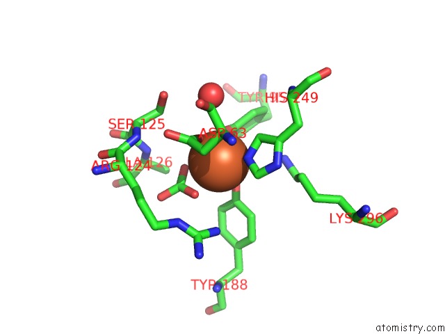
Mono view
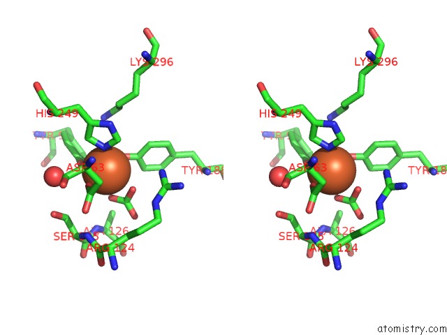
Stereo pair view
Mono view
Stereo pair view
A full contact list of Iron with other atoms in the Fe binding
site number 1 of Crystal Structure of Human Serum Transferrin, N-Lobe L66W Mutant within 5.0Å range:
|
Reference:
T.E.Adams,
A.B.Mason,
Q.Y.He,
P.J.Halbrooks,
S.K.Briggs,
V.C.Smith,
R.T.Macgillivray,
S.J.Everse.
The Position of Arginine 124 Controls the Rate of Iron Release From the N-Lobe of Human Serum Transferrin. A Structural Study J.Biol.Chem. V. 278 6027 2003.
ISSN: ISSN 0021-9258
PubMed: 12458193
DOI: 10.1074/JBC.M210349200
Page generated: Wed Jul 16 18:31:01 2025
ISSN: ISSN 0021-9258
PubMed: 12458193
DOI: 10.1074/JBC.M210349200
Last articles
Fe in 2YXOFe in 2YRS
Fe in 2YXC
Fe in 2YNM
Fe in 2YVJ
Fe in 2YP1
Fe in 2YU2
Fe in 2YU1
Fe in 2YQB
Fe in 2YOO