Iron »
PDB 1n5w-1nmi »
1nej »
Iron in PDB 1nej: Crystalline Human Carbonmonoxy Hemoglobin S (Liganded Sickle Cell Hemoglobin) Exhibits the R2 Quaternary State at Neutral pH in the Presence of Polyethylene Glycol: the 2.1 Angstrom Resolution Crystal Structure
Protein crystallography data
The structure of Crystalline Human Carbonmonoxy Hemoglobin S (Liganded Sickle Cell Hemoglobin) Exhibits the R2 Quaternary State at Neutral pH in the Presence of Polyethylene Glycol: the 2.1 Angstrom Resolution Crystal Structure, PDB code: 1nej
was solved by
L.N.Patskovska,
Y.V.Patskovsky,
S.C.Almo,
R.E.Hirsch,
with X-Ray Crystallography technique. A brief refinement statistics is given in the table below:
| Resolution Low / High (Å) | 8.00 / 2.10 |
| Space group | P 21 21 21 |
| Cell size a, b, c (Å), α, β, γ (°) | 58.000, 58.530, 171.840, 90.00, 90.00, 90.00 |
| R / Rfree (%) | 23.5 / 26.8 |
Iron Binding Sites:
The binding sites of Iron atom in the Crystalline Human Carbonmonoxy Hemoglobin S (Liganded Sickle Cell Hemoglobin) Exhibits the R2 Quaternary State at Neutral pH in the Presence of Polyethylene Glycol: the 2.1 Angstrom Resolution Crystal Structure
(pdb code 1nej). This binding sites where shown within
5.0 Angstroms radius around Iron atom.
In total 4 binding sites of Iron where determined in the Crystalline Human Carbonmonoxy Hemoglobin S (Liganded Sickle Cell Hemoglobin) Exhibits the R2 Quaternary State at Neutral pH in the Presence of Polyethylene Glycol: the 2.1 Angstrom Resolution Crystal Structure, PDB code: 1nej:
Jump to Iron binding site number: 1; 2; 3; 4;
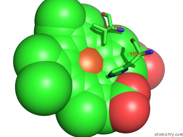
Iron binding site 1 out of 4 in 1nej
Go back to
Iron binding site 1 out
of 4 in the Crystalline Human Carbonmonoxy Hemoglobin S (Liganded Sickle Cell Hemoglobin) Exhibits the R2 Quaternary State at Neutral pH in the Presence of Polyethylene Glycol: the 2.1 Angstrom Resolution Crystal Structure
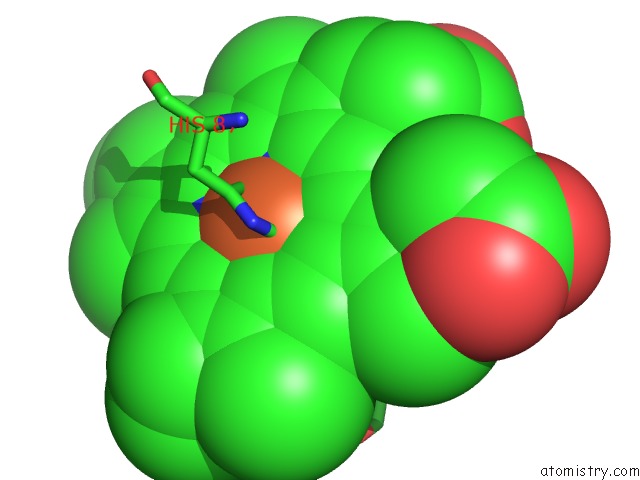
Mono view
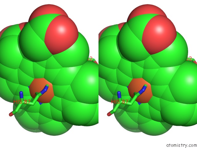
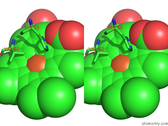
Stereo pair view
Mono view
Stereo pair view
A full contact list of Iron with other atoms in the Fe binding
site number 1 of Crystalline Human Carbonmonoxy Hemoglobin S (Liganded Sickle Cell Hemoglobin) Exhibits the R2 Quaternary State at Neutral pH in the Presence of Polyethylene Glycol: the 2.1 Angstrom Resolution Crystal Structure within 5.0Å range:
|
Iron binding site 2 out of 4 in 1nej
Go back to
Iron binding site 2 out
of 4 in the Crystalline Human Carbonmonoxy Hemoglobin S (Liganded Sickle Cell Hemoglobin) Exhibits the R2 Quaternary State at Neutral pH in the Presence of Polyethylene Glycol: the 2.1 Angstrom Resolution Crystal Structure
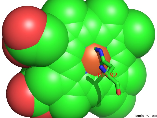
Mono view
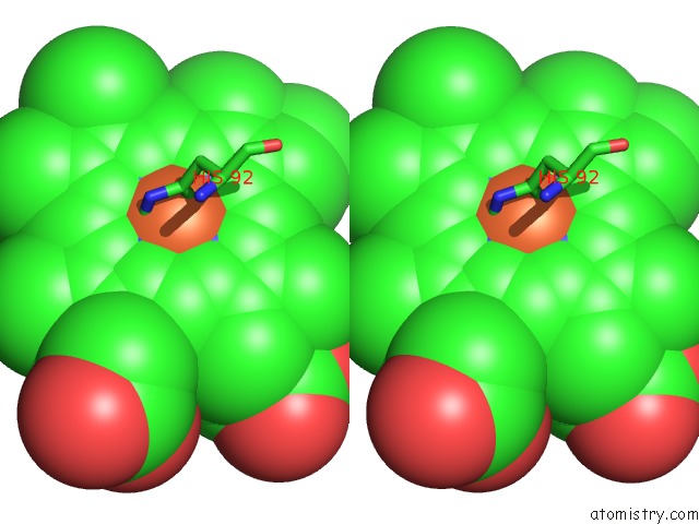
Stereo pair view
Mono view
Stereo pair view
A full contact list of Iron with other atoms in the Fe binding
site number 2 of Crystalline Human Carbonmonoxy Hemoglobin S (Liganded Sickle Cell Hemoglobin) Exhibits the R2 Quaternary State at Neutral pH in the Presence of Polyethylene Glycol: the 2.1 Angstrom Resolution Crystal Structure within 5.0Å range:
|
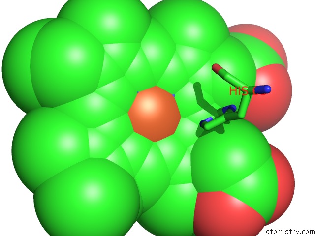
Iron binding site 3 out of 4 in 1nej
Go back to
Iron binding site 3 out
of 4 in the Crystalline Human Carbonmonoxy Hemoglobin S (Liganded Sickle Cell Hemoglobin) Exhibits the R2 Quaternary State at Neutral pH in the Presence of Polyethylene Glycol: the 2.1 Angstrom Resolution Crystal Structure
Mono view
Stereo pair view
Mono view
Stereo pair view
A full contact list of Iron with other atoms in the Fe binding
site number 3 of Crystalline Human Carbonmonoxy Hemoglobin S (Liganded Sickle Cell Hemoglobin) Exhibits the R2 Quaternary State at Neutral pH in the Presence of Polyethylene Glycol: the 2.1 Angstrom Resolution Crystal Structure within 5.0Å range:
|
Iron binding site 4 out of 4 in 1nej
Go back to
Iron binding site 4 out
of 4 in the Crystalline Human Carbonmonoxy Hemoglobin S (Liganded Sickle Cell Hemoglobin) Exhibits the R2 Quaternary State at Neutral pH in the Presence of Polyethylene Glycol: the 2.1 Angstrom Resolution Crystal Structure
Mono view
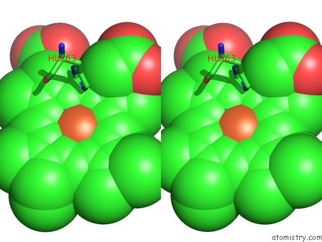
Stereo pair view
Mono view
Stereo pair view
A full contact list of Iron with other atoms in the Fe binding
site number 4 of Crystalline Human Carbonmonoxy Hemoglobin S (Liganded Sickle Cell Hemoglobin) Exhibits the R2 Quaternary State at Neutral pH in the Presence of Polyethylene Glycol: the 2.1 Angstrom Resolution Crystal Structure within 5.0Å range:
|
Reference:
L.N.Patskovska,
Y.V.Patskovsky,
S.C.Almo,
R.E.Hirsch.
Cohbc and Cohbs Crystallize in the R2 Quaternary State at Neutral pH in the Presence of Peg 4000. Acta Crystallogr.,Sect.D V. 61 566 2005.
ISSN: ISSN 0907-4449
PubMed: 15858266
DOI: 10.1107/S0907444905004622
Page generated: Wed Jul 16 18:33:38 2025
ISSN: ISSN 0907-4449
PubMed: 15858266
DOI: 10.1107/S0907444905004622
Last articles
I in 7DCZI in 7D36
I in 7D2V
I in 7D5A
I in 7D2X
I in 7CX9
I in 7CP7
I in 7CRF
I in 7CP6
I in 7BYD