Iron »
PDB 1n5w-1nmi »
1nip »
Iron in PDB 1nip: Crystallographic Structure of the Nitrogenase Iron Protein From Azotobacter Vinelandii
Protein crystallography data
The structure of Crystallographic Structure of the Nitrogenase Iron Protein From Azotobacter Vinelandii, PDB code: 1nip
was solved by
H.Komiya,
M.M.Georgiadis,
P.Chakrabarti,
D.Woo,
J.J.Kornuc,
D.C.Rees,
with X-Ray Crystallography technique. A brief refinement statistics is given in the table below:
| Resolution Low / High (Å) | 8.00 / 2.90 |
| Space group | P 1 21 1 |
| Cell size a, b, c (Å), α, β, γ (°) | 56.800, 92.900, 63.600, 90.00, 100.00, 90.00 |
| R / Rfree (%) | n/a / n/a |
Other elements in 1nip:
The structure of Crystallographic Structure of the Nitrogenase Iron Protein From Azotobacter Vinelandii also contains other interesting chemical elements:
| Magnesium | (Mg) | 2 atoms |
Iron Binding Sites:
The binding sites of Iron atom in the Crystallographic Structure of the Nitrogenase Iron Protein From Azotobacter Vinelandii
(pdb code 1nip). This binding sites where shown within
5.0 Angstroms radius around Iron atom.
In total 4 binding sites of Iron where determined in the Crystallographic Structure of the Nitrogenase Iron Protein From Azotobacter Vinelandii, PDB code: 1nip:
Jump to Iron binding site number: 1; 2; 3; 4;
In total 4 binding sites of Iron where determined in the Crystallographic Structure of the Nitrogenase Iron Protein From Azotobacter Vinelandii, PDB code: 1nip:
Jump to Iron binding site number: 1; 2; 3; 4;
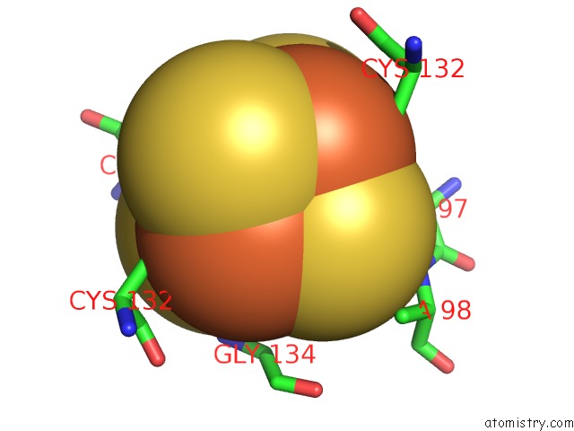
Iron binding site 1 out of 4 in 1nip
Go back to
Iron binding site 1 out
of 4 in the Crystallographic Structure of the Nitrogenase Iron Protein From Azotobacter Vinelandii
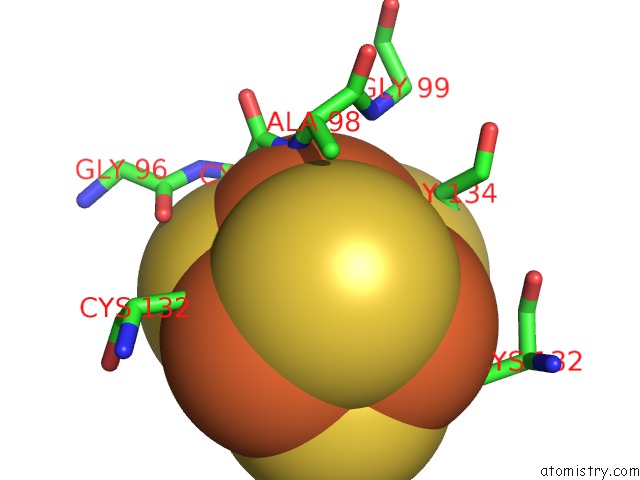
Mono view
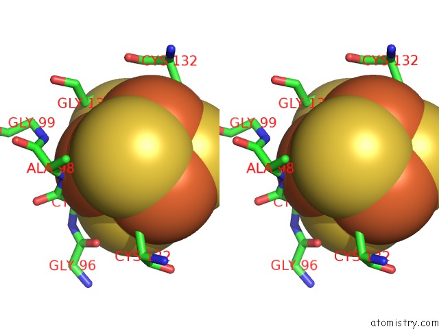
Stereo pair view
Mono view
Stereo pair view
|
|
A full contact list of Iron with other atoms in the Fe binding
site number 1 of Crystallographic Structure of the Nitrogenase Iron Protein From Azotobacter Vinelandii within 5.0Å range:
|
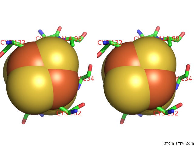
Iron binding site 2 out of 4 in 1nip
Go back to
Iron binding site 2 out
of 4 in the Crystallographic Structure of the Nitrogenase Iron Protein From Azotobacter Vinelandii
Mono view
Stereo pair view
Mono view
Stereo pair view
|
|
A full contact list of Iron with other atoms in the Fe binding
site number 2 of Crystallographic Structure of the Nitrogenase Iron Protein From Azotobacter Vinelandii within 5.0Å range:
|
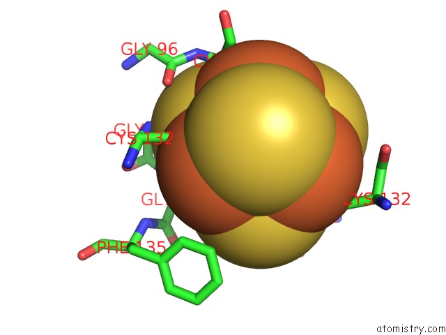
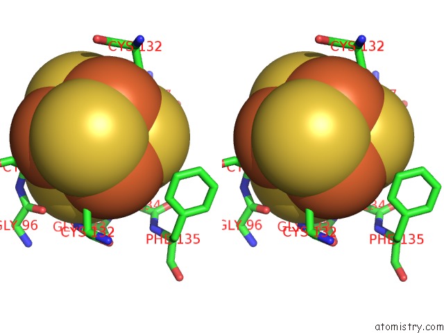
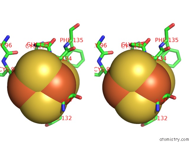
Iron binding site 3 out of 4 in 1nip
Go back to
Iron binding site 3 out
of 4 in the Crystallographic Structure of the Nitrogenase Iron Protein From Azotobacter Vinelandii
Mono view
Stereo pair view
Mono view
Stereo pair view
|
|
A full contact list of Iron with other atoms in the Fe binding
site number 3 of Crystallographic Structure of the Nitrogenase Iron Protein From Azotobacter Vinelandii within 5.0Å range:
|
Iron binding site 4 out of 4 in 1nip
Go back to
Iron binding site 4 out
of 4 in the Crystallographic Structure of the Nitrogenase Iron Protein From Azotobacter Vinelandii

Mono view
Stereo pair view
Mono view
Stereo pair view
|
|
A full contact list of Iron with other atoms in the Fe binding
site number 4 of Crystallographic Structure of the Nitrogenase Iron Protein From Azotobacter Vinelandii within 5.0Å range:
|
Reference:
M.M.Georgiadis,
H.Komiya,
P.Chakrabarti,
D.Woo,
J.J.Kornuc,
D.C.Rees.
Crystallographic Structure of the Nitrogenase Iron Protein From Azotobacter Vinelandii. Science V. 257 1653 1992.
ISSN: ISSN 0036-8075
PubMed: 1529353
Page generated: Wed Jul 16 18:42:22 2025
ISSN: ISSN 0036-8075
PubMed: 1529353
Last articles
Mg in 7JYYMg in 7JY8
Mg in 7JYV
Mg in 7JY7
Mg in 7JYU
Mg in 7JY6
Mg in 7JY5
Mg in 7JXQ
Mg in 7JXP
Mg in 7JXM