Iron »
PDB 1nml-1o1i »
1nr6 »
Iron in PDB 1nr6: Microsomal Cytochrome P450 2C5/3LVDH Complex with Diclofenac
Enzymatic activity of Microsomal Cytochrome P450 2C5/3LVDH Complex with Diclofenac
All present enzymatic activity of Microsomal Cytochrome P450 2C5/3LVDH Complex with Diclofenac:
1.14.14.1;
1.14.14.1;
Protein crystallography data
The structure of Microsomal Cytochrome P450 2C5/3LVDH Complex with Diclofenac, PDB code: 1nr6
was solved by
M.R.Wester,
E.F.Johnson,
C.D.Stout,
with X-Ray Crystallography technique. A brief refinement statistics is given in the table below:
| Resolution Low / High (Å) | 50.00 / 2.10 |
| Space group | I 2 2 2 |
| Cell size a, b, c (Å), α, β, γ (°) | 73.940, 130.020, 172.780, 90.00, 90.00, 90.00 |
| R / Rfree (%) | 23.1 / 26.8 |
Other elements in 1nr6:
The structure of Microsomal Cytochrome P450 2C5/3LVDH Complex with Diclofenac also contains other interesting chemical elements:
| Chlorine | (Cl) | 2 atoms |
Iron Binding Sites:
The binding sites of Iron atom in the Microsomal Cytochrome P450 2C5/3LVDH Complex with Diclofenac
(pdb code 1nr6). This binding sites where shown within
5.0 Angstroms radius around Iron atom.
In total only one binding site of Iron was determined in the Microsomal Cytochrome P450 2C5/3LVDH Complex with Diclofenac, PDB code: 1nr6:
In total only one binding site of Iron was determined in the Microsomal Cytochrome P450 2C5/3LVDH Complex with Diclofenac, PDB code: 1nr6:
Iron binding site 1 out of 1 in 1nr6
Go back to
Iron binding site 1 out
of 1 in the Microsomal Cytochrome P450 2C5/3LVDH Complex with Diclofenac
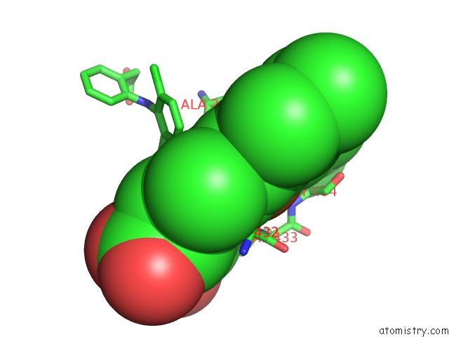
Mono view
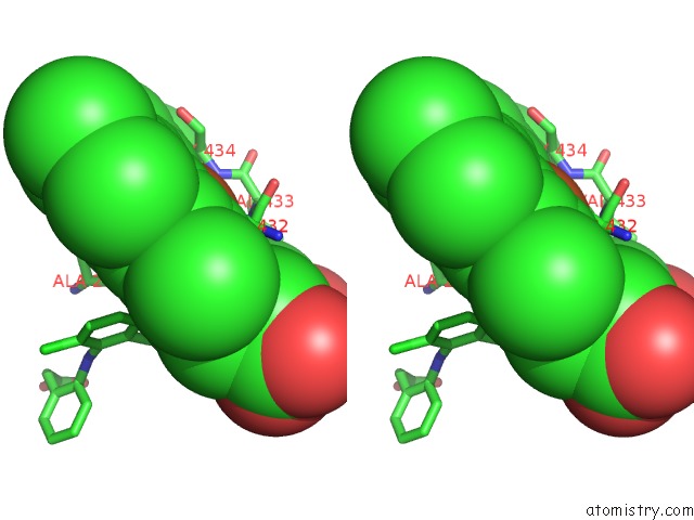
Stereo pair view
Mono view
Stereo pair view
A full contact list of Iron with other atoms in the Fe binding
site number 1 of Microsomal Cytochrome P450 2C5/3LVDH Complex with Diclofenac within 5.0Å range:
|
Reference:
M.R.Wester,
E.F.Johnson,
C.Marques-Soares,
S.Dijols,
P.M.Dansette,
D.Mansuy,
C.D.Stout.
Structure of Mammalian Cytochrome P450 2C5 Complexed with Diclofenac at 2.1 A Resolution: Evidence For An Induced Fit Model of Substrate Binding Biochemistry V. 42 9335 2003.
ISSN: ISSN 0006-2960
PubMed: 12899620
DOI: 10.1021/BI034556L
Page generated: Wed Jul 16 18:52:14 2025
ISSN: ISSN 0006-2960
PubMed: 12899620
DOI: 10.1021/BI034556L
Last articles
Mg in 1C5PMg in 1C2M
Mg in 1C2J
Mg in 1C2H
Mg in 1C2G
Mg in 1BZY
Mg in 1C2F
Mg in 1C2E
Mg in 1C2D
Mg in 1C1T