Iron »
PDB 1nml-1o1i »
1ns6 »
Iron in PDB 1ns6: The 2.1A Structure of Horse (Alpha Hemichrome/Beta Met) Hemoglobin at pH 5.4
Protein crystallography data
The structure of The 2.1A Structure of Horse (Alpha Hemichrome/Beta Met) Hemoglobin at pH 5.4, PDB code: 1ns6
was solved by
V.L.Robinson,
B.B.Smith,
A.Arnone,
with X-Ray Crystallography technique. A brief refinement statistics is given in the table below:
| Resolution Low / High (Å) | 54.23 / 2.05 |
| Space group | C 1 2 1 |
| Cell size a, b, c (Å), α, β, γ (°) | 108.110, 63.330, 53.850, 90.00, 84.72, 90.00 |
| R / Rfree (%) | 16.3 / 18.8 |
Iron Binding Sites:
The binding sites of Iron atom in the The 2.1A Structure of Horse (Alpha Hemichrome/Beta Met) Hemoglobin at pH 5.4
(pdb code 1ns6). This binding sites where shown within
5.0 Angstroms radius around Iron atom.
In total 2 binding sites of Iron where determined in the The 2.1A Structure of Horse (Alpha Hemichrome/Beta Met) Hemoglobin at pH 5.4, PDB code: 1ns6:
Jump to Iron binding site number: 1; 2;
In total 2 binding sites of Iron where determined in the The 2.1A Structure of Horse (Alpha Hemichrome/Beta Met) Hemoglobin at pH 5.4, PDB code: 1ns6:
Jump to Iron binding site number: 1; 2;
Iron binding site 1 out of 2 in 1ns6
Go back to
Iron binding site 1 out
of 2 in the The 2.1A Structure of Horse (Alpha Hemichrome/Beta Met) Hemoglobin at pH 5.4
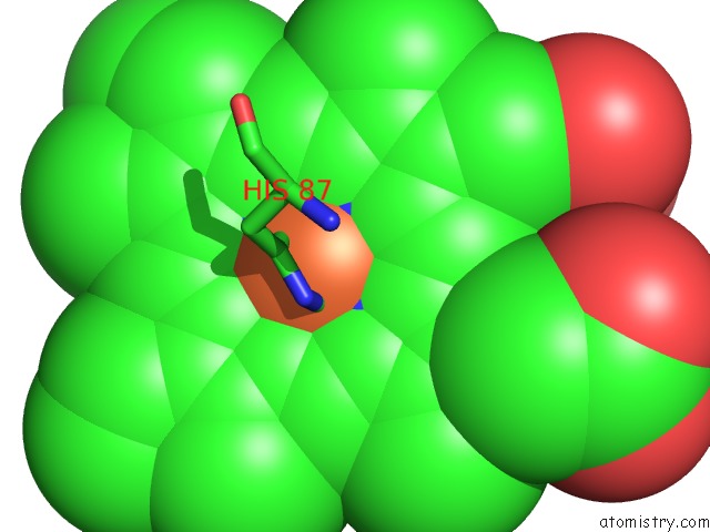
Mono view
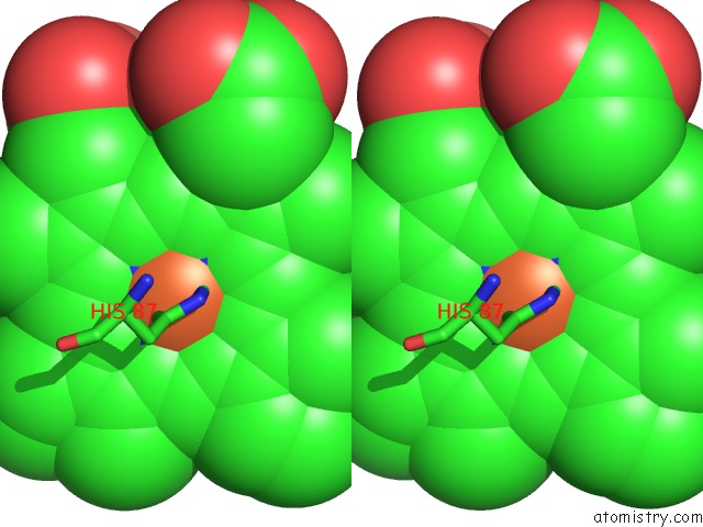
Stereo pair view
Mono view
Stereo pair view
A full contact list of Iron with other atoms in the Fe binding
site number 1 of The 2.1A Structure of Horse (Alpha Hemichrome/Beta Met) Hemoglobin at pH 5.4 within 5.0Å range:
|
Iron binding site 2 out of 2 in 1ns6
Go back to
Iron binding site 2 out
of 2 in the The 2.1A Structure of Horse (Alpha Hemichrome/Beta Met) Hemoglobin at pH 5.4
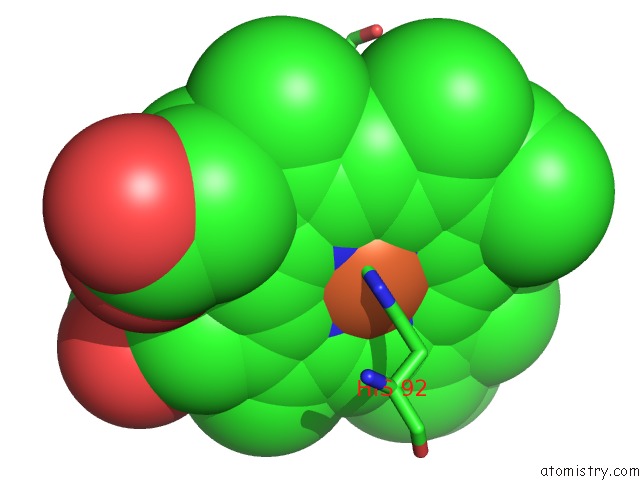
Mono view
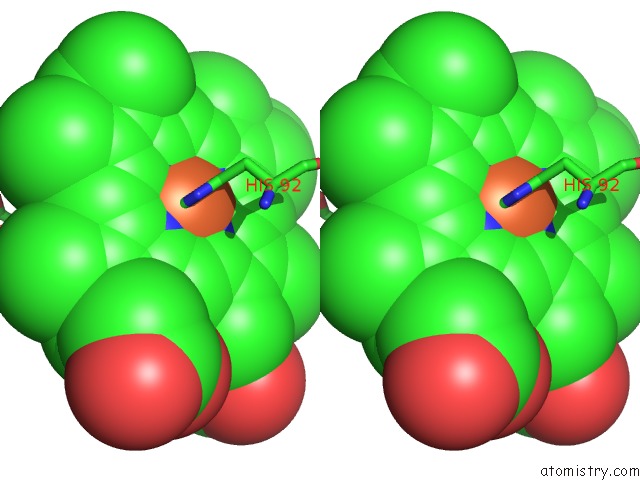
Stereo pair view
Mono view
Stereo pair view
A full contact list of Iron with other atoms in the Fe binding
site number 2 of The 2.1A Structure of Horse (Alpha Hemichrome/Beta Met) Hemoglobin at pH 5.4 within 5.0Å range:
|
Reference:
V.L.Robinson,
B.B.Smith,
A.Arnone.
A pH-Dependent Aquomet-to-Hemichrome Transition in Crystalline Horse Methemoglobin Biochemistry V. 42 10113 2003.
ISSN: ISSN 0006-2960
PubMed: 12939139
DOI: 10.1021/BI030059T
Page generated: Wed Jul 16 18:52:30 2025
ISSN: ISSN 0006-2960
PubMed: 12939139
DOI: 10.1021/BI030059T
Last articles
Mg in 4QBYMg in 4QF5
Mg in 4QFM
Mg in 4QEH
Mg in 4QEL
Mg in 4QE5
Mg in 4QDG
Mg in 4QDM
Mg in 4QDI
Mg in 4QBL