Iron »
PDB 1o1j-1off »
1obm »
Iron in PDB 1obm: Recombinant Sperm Whale Myoglobin 29F/64Q/68F/122N Mutant (Met)
Protein crystallography data
The structure of Recombinant Sperm Whale Myoglobin 29F/64Q/68F/122N Mutant (Met), PDB code: 1obm
was solved by
E.A.Brucker,
R.A.Lile,
G.N.Phillips Jr.,
with X-Ray Crystallography technique. A brief refinement statistics is given in the table below:
| Resolution Low / High (Å) | 5.00 / 1.85 |
| Space group | P 6 |
| Cell size a, b, c (Å), α, β, γ (°) | 91.390, 91.390, 45.750, 90.00, 90.00, 120.00 |
| R / Rfree (%) | 16.8 / n/a |
Iron Binding Sites:
The binding sites of Iron atom in the Recombinant Sperm Whale Myoglobin 29F/64Q/68F/122N Mutant (Met)
(pdb code 1obm). This binding sites where shown within
5.0 Angstroms radius around Iron atom.
In total only one binding site of Iron was determined in the Recombinant Sperm Whale Myoglobin 29F/64Q/68F/122N Mutant (Met), PDB code: 1obm:
In total only one binding site of Iron was determined in the Recombinant Sperm Whale Myoglobin 29F/64Q/68F/122N Mutant (Met), PDB code: 1obm:
Iron binding site 1 out of 1 in 1obm
Go back to
Iron binding site 1 out
of 1 in the Recombinant Sperm Whale Myoglobin 29F/64Q/68F/122N Mutant (Met)
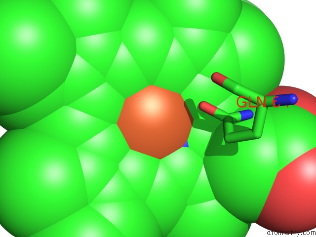
Mono view
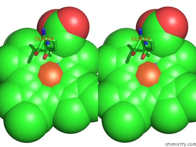
Stereo pair view
Mono view
Stereo pair view
A full contact list of Iron with other atoms in the Fe binding
site number 1 of Recombinant Sperm Whale Myoglobin 29F/64Q/68F/122N Mutant (Met) within 5.0Å range:
|
Reference:
B.D.Nguyen,
X.Zhao,
K.Vyas,
G.N.La Mar,
R.A.Lile,
E.A.Brucker,
G.N.Phillips Jr.,
J.S.Olson,
J.B.Wittenberg.
Solution and Crystal Structures of A Sperm Whale Myoglobin Triple Mutant That Mimics the Sulfide-Binding Hemoglobin From Lucina Pectinata. J.Biol.Chem. V. 273 9517 1998.
ISSN: ISSN 0021-9258
PubMed: 9545280
DOI: 10.1074/JBC.273.16.9517
Page generated: Wed Jul 16 19:05:34 2025
ISSN: ISSN 0021-9258
PubMed: 9545280
DOI: 10.1074/JBC.273.16.9517
Last articles
Ni in 1EM0Ni in 1ELW
Ni in 1EJX
Ni in 1ELR
Ni in 1EK0
Ni in 1EJW
Ni in 1EJV
Ni in 1EJU
Ni in 1EJT
Ni in 1EJS