Iron »
PDB 1ofj-1ozl »
1oqb »
Iron in PDB 1oqb: The Crystal Structure of the One-Iron Form of the Di-Iron Center in Stearoyl Acyl Carrier Protein Desaturase From Ricinus Communis (Castor Bean).
Enzymatic activity of The Crystal Structure of the One-Iron Form of the Di-Iron Center in Stearoyl Acyl Carrier Protein Desaturase From Ricinus Communis (Castor Bean).
All present enzymatic activity of The Crystal Structure of the One-Iron Form of the Di-Iron Center in Stearoyl Acyl Carrier Protein Desaturase From Ricinus Communis (Castor Bean).:
1.14.19.2;
1.14.19.2;
Protein crystallography data
The structure of The Crystal Structure of the One-Iron Form of the Di-Iron Center in Stearoyl Acyl Carrier Protein Desaturase From Ricinus Communis (Castor Bean)., PDB code: 1oqb
was solved by
M.Moche,
J.Shanklin,
A.K.Ghoshal,
Y.Lindqvist,
with X-Ray Crystallography technique. A brief refinement statistics is given in the table below:
| Resolution Low / High (Å) | 20.00 / 2.80 |
| Space group | P 21 21 21 |
| Cell size a, b, c (Å), α, β, γ (°) | 81.908, 145.791, 192.424, 90.00, 90.00, 90.00 |
| R / Rfree (%) | 22.4 / 24.8 |
Iron Binding Sites:
The binding sites of Iron atom in the The Crystal Structure of the One-Iron Form of the Di-Iron Center in Stearoyl Acyl Carrier Protein Desaturase From Ricinus Communis (Castor Bean).
(pdb code 1oqb). This binding sites where shown within
5.0 Angstroms radius around Iron atom.
In total 6 binding sites of Iron where determined in the The Crystal Structure of the One-Iron Form of the Di-Iron Center in Stearoyl Acyl Carrier Protein Desaturase From Ricinus Communis (Castor Bean)., PDB code: 1oqb:
Jump to Iron binding site number: 1; 2; 3; 4; 5; 6;
In total 6 binding sites of Iron where determined in the The Crystal Structure of the One-Iron Form of the Di-Iron Center in Stearoyl Acyl Carrier Protein Desaturase From Ricinus Communis (Castor Bean)., PDB code: 1oqb:
Jump to Iron binding site number: 1; 2; 3; 4; 5; 6;
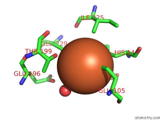
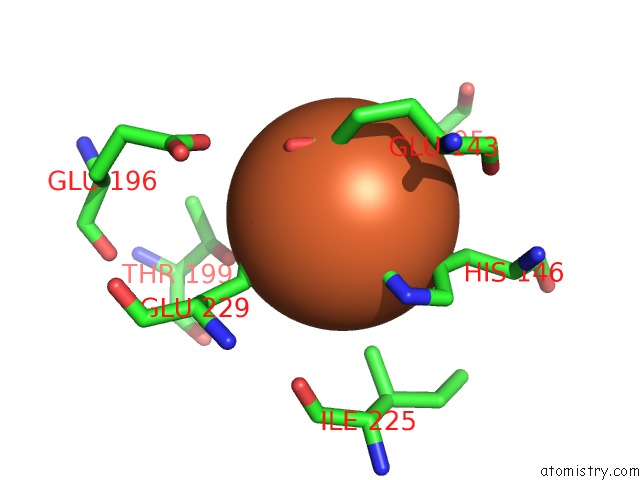
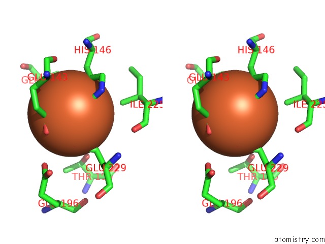
Iron binding site 1 out of 6 in 1oqb
Go back to
Iron binding site 1 out
of 6 in the The Crystal Structure of the One-Iron Form of the Di-Iron Center in Stearoyl Acyl Carrier Protein Desaturase From Ricinus Communis (Castor Bean).
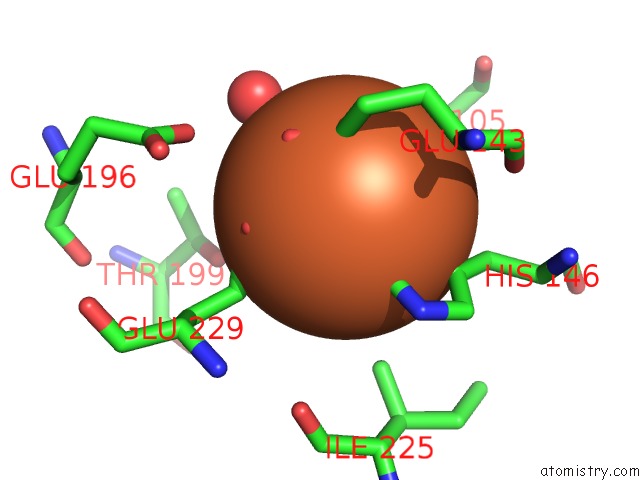
Mono view
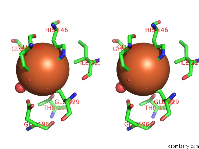
Stereo pair view
Mono view
Stereo pair view
A full contact list of Iron with other atoms in the Fe binding
site number 1 of The Crystal Structure of the One-Iron Form of the Di-Iron Center in Stearoyl Acyl Carrier Protein Desaturase From Ricinus Communis (Castor Bean). within 5.0Å range:
|
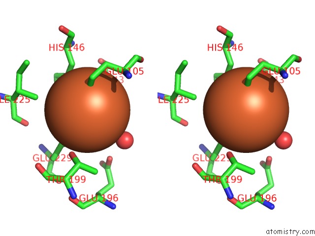
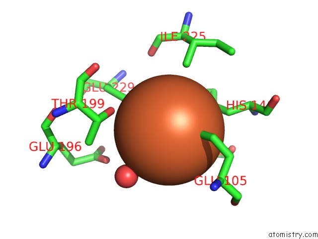
Iron binding site 2 out of 6 in 1oqb
Go back to
Iron binding site 2 out
of 6 in the The Crystal Structure of the One-Iron Form of the Di-Iron Center in Stearoyl Acyl Carrier Protein Desaturase From Ricinus Communis (Castor Bean).
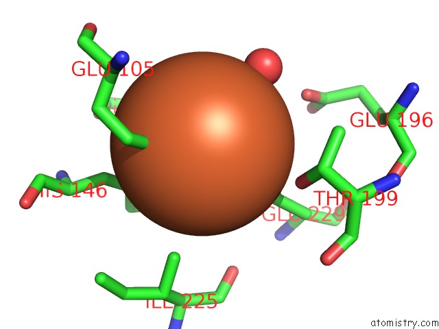
Mono view
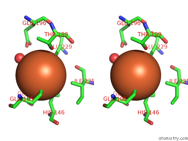
Stereo pair view
Mono view
Stereo pair view
A full contact list of Iron with other atoms in the Fe binding
site number 2 of The Crystal Structure of the One-Iron Form of the Di-Iron Center in Stearoyl Acyl Carrier Protein Desaturase From Ricinus Communis (Castor Bean). within 5.0Å range:
|
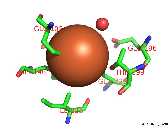
Iron binding site 3 out of 6 in 1oqb
Go back to
Iron binding site 3 out
of 6 in the The Crystal Structure of the One-Iron Form of the Di-Iron Center in Stearoyl Acyl Carrier Protein Desaturase From Ricinus Communis (Castor Bean).
Mono view
Stereo pair view
Mono view
Stereo pair view
A full contact list of Iron with other atoms in the Fe binding
site number 3 of The Crystal Structure of the One-Iron Form of the Di-Iron Center in Stearoyl Acyl Carrier Protein Desaturase From Ricinus Communis (Castor Bean). within 5.0Å range:
|
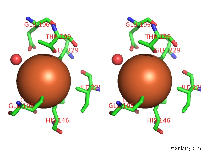
Iron binding site 4 out of 6 in 1oqb
Go back to
Iron binding site 4 out
of 6 in the The Crystal Structure of the One-Iron Form of the Di-Iron Center in Stearoyl Acyl Carrier Protein Desaturase From Ricinus Communis (Castor Bean).
Mono view
Stereo pair view
Mono view
Stereo pair view
A full contact list of Iron with other atoms in the Fe binding
site number 4 of The Crystal Structure of the One-Iron Form of the Di-Iron Center in Stearoyl Acyl Carrier Protein Desaturase From Ricinus Communis (Castor Bean). within 5.0Å range:
|
Iron binding site 5 out of 6 in 1oqb
Go back to
Iron binding site 5 out
of 6 in the The Crystal Structure of the One-Iron Form of the Di-Iron Center in Stearoyl Acyl Carrier Protein Desaturase From Ricinus Communis (Castor Bean).
Mono view
Stereo pair view
Mono view
Stereo pair view
A full contact list of Iron with other atoms in the Fe binding
site number 5 of The Crystal Structure of the One-Iron Form of the Di-Iron Center in Stearoyl Acyl Carrier Protein Desaturase From Ricinus Communis (Castor Bean). within 5.0Å range:
|
Iron binding site 6 out of 6 in 1oqb
Go back to
Iron binding site 6 out
of 6 in the The Crystal Structure of the One-Iron Form of the Di-Iron Center in Stearoyl Acyl Carrier Protein Desaturase From Ricinus Communis (Castor Bean).
Mono view

Stereo pair view
Mono view
Stereo pair view
A full contact list of Iron with other atoms in the Fe binding
site number 6 of The Crystal Structure of the One-Iron Form of the Di-Iron Center in Stearoyl Acyl Carrier Protein Desaturase From Ricinus Communis (Castor Bean). within 5.0Å range:
|
Reference:
M.Moche,
J.Shanklin,
A.Ghoshal,
Y.Lindqvist.
Azide and Acetate Complexes Plus Two Iron-Depleted Crystal Structures of the Di-Iron Enzyme DELTA9 Stearoyl-Acp Desaturase-Implications For Oxygen Activation and Catalytic Intermediates J.Biol.Chem. V. 278 25072 2003.
ISSN: ISSN 0021-9258
PubMed: 12704186
DOI: 10.1074/JBC.M301662200
Page generated: Wed Jul 16 19:19:19 2025
ISSN: ISSN 0021-9258
PubMed: 12704186
DOI: 10.1074/JBC.M301662200
Last articles
Na in 3MICNa in 3MI3
Na in 3MIE
Na in 3MGI
Na in 3ME4
Na in 3MDC
Na in 3MGA
Na in 3MEQ
Na in 3ME2
Na in 3MDA