Iron »
PDB 1ofj-1ozl »
1out »
Iron in PDB 1out: Trout Hemoglobin I
Protein crystallography data
The structure of Trout Hemoglobin I, PDB code: 1out
was solved by
J.Tame,
J.Wilson,
with X-Ray Crystallography technique. A brief refinement statistics is given in the table below:
| Resolution Low / High (Å) | 8.00 / 2.30 |
| Space group | P 65 2 2 |
| Cell size a, b, c (Å), α, β, γ (°) | 63.280, 63.280, 312.740, 90.00, 90.00, 120.00 |
| R / Rfree (%) | 16.2 / 24.7 |
Iron Binding Sites:
The binding sites of Iron atom in the Trout Hemoglobin I
(pdb code 1out). This binding sites where shown within
5.0 Angstroms radius around Iron atom.
In total 2 binding sites of Iron where determined in the Trout Hemoglobin I, PDB code: 1out:
Jump to Iron binding site number: 1; 2;
In total 2 binding sites of Iron where determined in the Trout Hemoglobin I, PDB code: 1out:
Jump to Iron binding site number: 1; 2;
Iron binding site 1 out of 2 in 1out
Go back to
Iron binding site 1 out
of 2 in the Trout Hemoglobin I
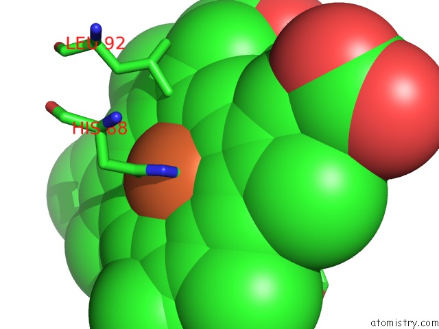
Mono view
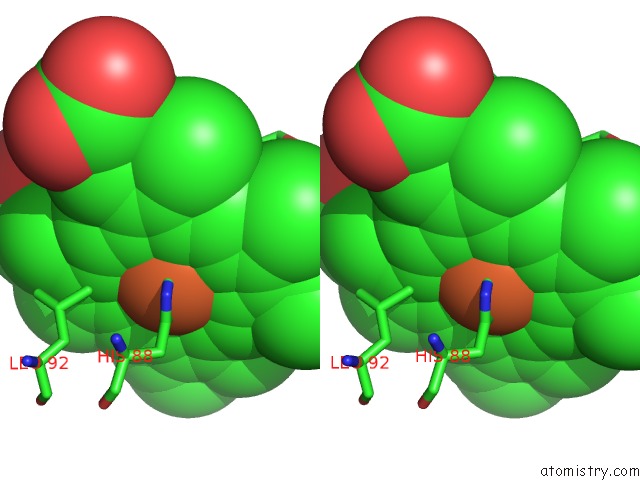
Stereo pair view
Mono view
Stereo pair view
A full contact list of Iron with other atoms in the Fe binding
site number 1 of Trout Hemoglobin I within 5.0Å range:
|
Iron binding site 2 out of 2 in 1out
Go back to
Iron binding site 2 out
of 2 in the Trout Hemoglobin I
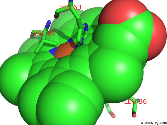
Mono view
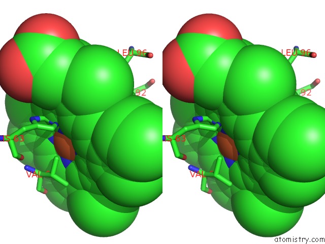
Stereo pair view
Mono view
Stereo pair view
A full contact list of Iron with other atoms in the Fe binding
site number 2 of Trout Hemoglobin I within 5.0Å range:
|
Reference:
J.R.Tame,
J.C.Wilson,
R.E.Weber.
The Crystal Structures of Trout Hb I in the Deoxy and Carbonmonoxy Forms. J.Mol.Biol. V. 259 749 1996.
ISSN: ISSN 0022-2836
PubMed: 8683580
DOI: 10.1006/JMBI.1996.0355
Page generated: Wed Jul 16 19:25:10 2025
ISSN: ISSN 0022-2836
PubMed: 8683580
DOI: 10.1006/JMBI.1996.0355
Last articles
Na in 3MICNa in 3MI3
Na in 3MIE
Na in 3MGI
Na in 3ME4
Na in 3MDC
Na in 3MGA
Na in 3MEQ
Na in 3ME2
Na in 3MDA