Iron »
PDB 1ozr-1pha »
1pby »
Iron in PDB 1pby: Structure of the Phenylhydrazine Adduct of the Quinohemoprotein Amine Dehydrogenase From Paracoccus Denitrificans at 1.7 A Resolution
Enzymatic activity of Structure of the Phenylhydrazine Adduct of the Quinohemoprotein Amine Dehydrogenase From Paracoccus Denitrificans at 1.7 A Resolution
All present enzymatic activity of Structure of the Phenylhydrazine Adduct of the Quinohemoprotein Amine Dehydrogenase From Paracoccus Denitrificans at 1.7 A Resolution:
1.4.99.3;
1.4.99.3;
Protein crystallography data
The structure of Structure of the Phenylhydrazine Adduct of the Quinohemoprotein Amine Dehydrogenase From Paracoccus Denitrificans at 1.7 A Resolution, PDB code: 1pby
was solved by
S.Datta,
T.Ikeda,
K.Kano,
F.S.Mathews,
with X-Ray Crystallography technique. A brief refinement statistics is given in the table below:
| Resolution Low / High (Å) | 36.42 / 1.70 |
| Space group | P 41 21 2 |
| Cell size a, b, c (Å), α, β, γ (°) | 99.237, 99.237, 213.056, 90.00, 90.00, 90.00 |
| R / Rfree (%) | 19.1 / 22.4 |
Iron Binding Sites:
The binding sites of Iron atom in the Structure of the Phenylhydrazine Adduct of the Quinohemoprotein Amine Dehydrogenase From Paracoccus Denitrificans at 1.7 A Resolution
(pdb code 1pby). This binding sites where shown within
5.0 Angstroms radius around Iron atom.
In total 2 binding sites of Iron where determined in the Structure of the Phenylhydrazine Adduct of the Quinohemoprotein Amine Dehydrogenase From Paracoccus Denitrificans at 1.7 A Resolution, PDB code: 1pby:
Jump to Iron binding site number: 1; 2;
In total 2 binding sites of Iron where determined in the Structure of the Phenylhydrazine Adduct of the Quinohemoprotein Amine Dehydrogenase From Paracoccus Denitrificans at 1.7 A Resolution, PDB code: 1pby:
Jump to Iron binding site number: 1; 2;
Iron binding site 1 out of 2 in 1pby
Go back to
Iron binding site 1 out
of 2 in the Structure of the Phenylhydrazine Adduct of the Quinohemoprotein Amine Dehydrogenase From Paracoccus Denitrificans at 1.7 A Resolution
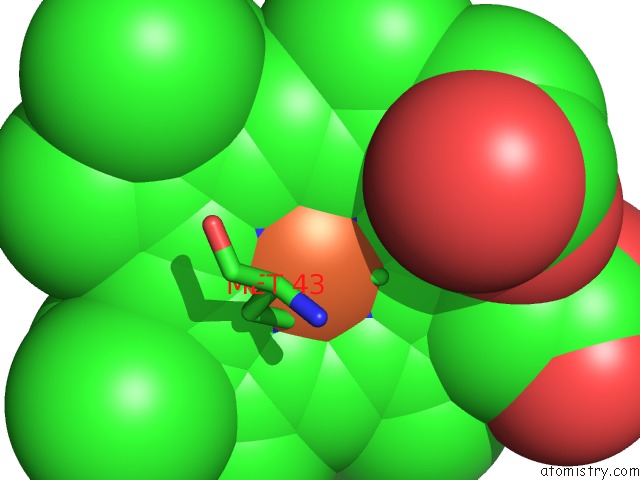
Mono view
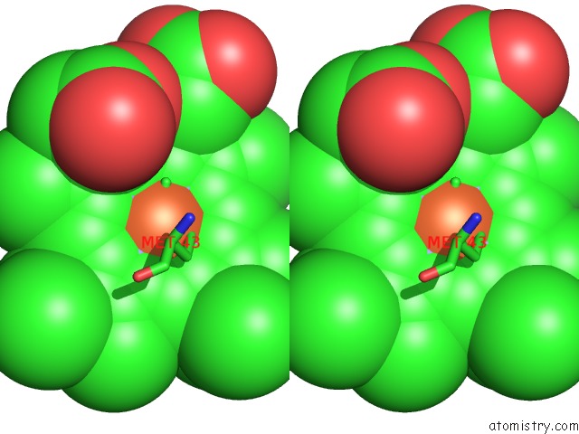
Stereo pair view
Mono view
Stereo pair view
A full contact list of Iron with other atoms in the Fe binding
site number 1 of Structure of the Phenylhydrazine Adduct of the Quinohemoprotein Amine Dehydrogenase From Paracoccus Denitrificans at 1.7 A Resolution within 5.0Å range:
|
Iron binding site 2 out of 2 in 1pby
Go back to
Iron binding site 2 out
of 2 in the Structure of the Phenylhydrazine Adduct of the Quinohemoprotein Amine Dehydrogenase From Paracoccus Denitrificans at 1.7 A Resolution
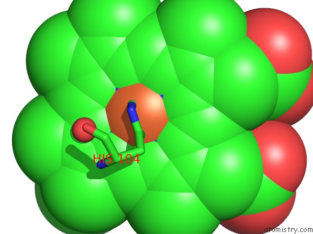
Mono view
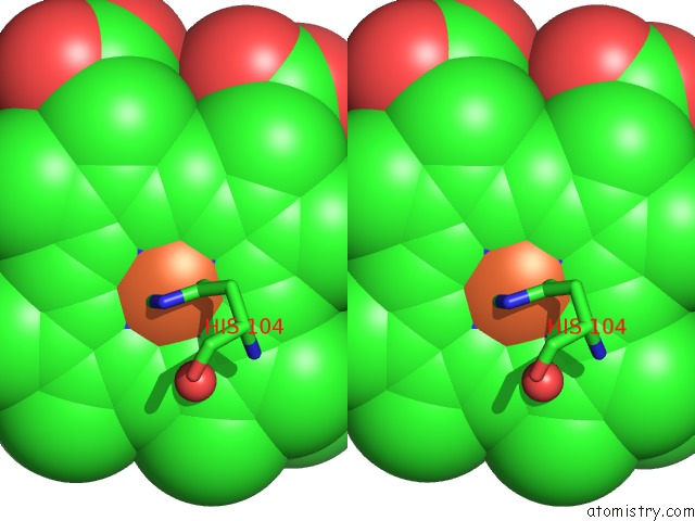
Stereo pair view
Mono view
Stereo pair view
A full contact list of Iron with other atoms in the Fe binding
site number 2 of Structure of the Phenylhydrazine Adduct of the Quinohemoprotein Amine Dehydrogenase From Paracoccus Denitrificans at 1.7 A Resolution within 5.0Å range:
|
Reference:
S.Datta,
T.Ikeda,
K.Kano,
F.S.Mathews.
Structure of the Phenylhydrazine Adduct of the Quinohemoprotein Amine Dehydrogenase From Paracoccus Denitrificans at 1.7 A Resolution. Acta Crystallogr.,Sect.D V. 59 1551 2003.
ISSN: ISSN 0907-4449
PubMed: 12925784
DOI: 10.1107/S090744490301429X
Page generated: Wed Jul 16 19:33:15 2025
ISSN: ISSN 0907-4449
PubMed: 12925784
DOI: 10.1107/S090744490301429X
Last articles
Fe in 2YXOFe in 2YRS
Fe in 2YXC
Fe in 2YNM
Fe in 2YVJ
Fe in 2YP1
Fe in 2YU2
Fe in 2YU1
Fe in 2YQB
Fe in 2YOO