Iron »
PDB 1phb-1q5d »
1q0o »
Iron in PDB 1q0o: Crystal Structure of Homoprotocatechuate 2,3-Dioxygenase From Brevibacterium Fuscum (Full Length Protein)
Enzymatic activity of Crystal Structure of Homoprotocatechuate 2,3-Dioxygenase From Brevibacterium Fuscum (Full Length Protein)
All present enzymatic activity of Crystal Structure of Homoprotocatechuate 2,3-Dioxygenase From Brevibacterium Fuscum (Full Length Protein):
1.13.11.15;
1.13.11.15;
Protein crystallography data
The structure of Crystal Structure of Homoprotocatechuate 2,3-Dioxygenase From Brevibacterium Fuscum (Full Length Protein), PDB code: 1q0o
was solved by
M.W.Vetting,
L.P.Wackett,
L.Que,
J.D.Lipscomb,
D.H.Ohlendorf,
with X-Ray Crystallography technique. A brief refinement statistics is given in the table below:
| Resolution Low / High (Å) | 20.00 / 2.30 |
| Space group | P 32 2 1 |
| Cell size a, b, c (Å), α, β, γ (°) | 118.900, 118.900, 110.300, 90.00, 90.00, 120.00 |
| R / Rfree (%) | 16.1 / 20.8 |
Iron Binding Sites:
The binding sites of Iron atom in the Crystal Structure of Homoprotocatechuate 2,3-Dioxygenase From Brevibacterium Fuscum (Full Length Protein)
(pdb code 1q0o). This binding sites where shown within
5.0 Angstroms radius around Iron atom.
In total 2 binding sites of Iron where determined in the Crystal Structure of Homoprotocatechuate 2,3-Dioxygenase From Brevibacterium Fuscum (Full Length Protein), PDB code: 1q0o:
Jump to Iron binding site number: 1; 2;
In total 2 binding sites of Iron where determined in the Crystal Structure of Homoprotocatechuate 2,3-Dioxygenase From Brevibacterium Fuscum (Full Length Protein), PDB code: 1q0o:
Jump to Iron binding site number: 1; 2;
Iron binding site 1 out of 2 in 1q0o
Go back to
Iron binding site 1 out
of 2 in the Crystal Structure of Homoprotocatechuate 2,3-Dioxygenase From Brevibacterium Fuscum (Full Length Protein)
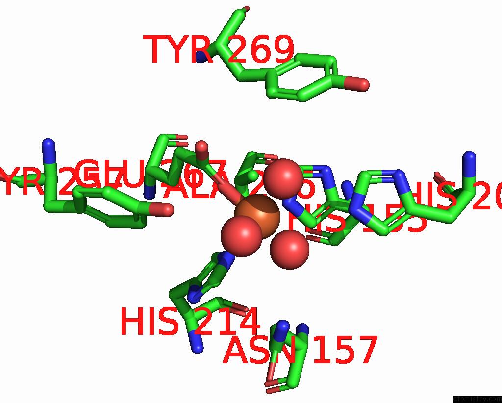
Mono view
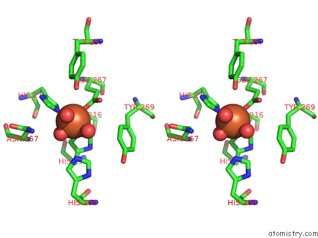
Stereo pair view
Mono view
Stereo pair view
A full contact list of Iron with other atoms in the Fe binding
site number 1 of Crystal Structure of Homoprotocatechuate 2,3-Dioxygenase From Brevibacterium Fuscum (Full Length Protein) within 5.0Å range:
|
Iron binding site 2 out of 2 in 1q0o
Go back to
Iron binding site 2 out
of 2 in the Crystal Structure of Homoprotocatechuate 2,3-Dioxygenase From Brevibacterium Fuscum (Full Length Protein)
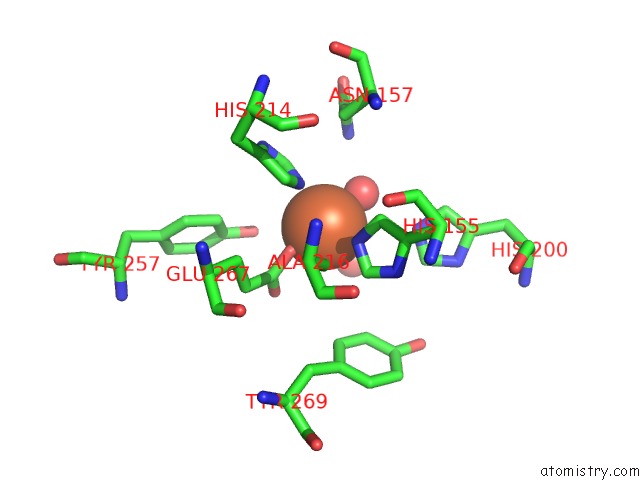
Mono view
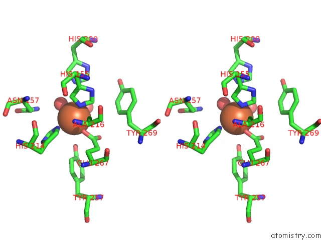
Stereo pair view
Mono view
Stereo pair view
A full contact list of Iron with other atoms in the Fe binding
site number 2 of Crystal Structure of Homoprotocatechuate 2,3-Dioxygenase From Brevibacterium Fuscum (Full Length Protein) within 5.0Å range:
|
Reference:
M.W.Vetting,
L.P.Wackett,
L.Que,
J.D.Lipscomb,
D.H.Ohlendorf.
Crystallographic Comparison of Manganese- and Iron-Dependent Homoprotocatechuate 2,3-Dioxygenases. J.Bacteriol. V. 186 1945 2004.
ISSN: ISSN 0021-9193
PubMed: 15028678
DOI: 10.1128/JB.186.7.1945-1958.2004
Page generated: Wed Jul 16 19:43:35 2025
ISSN: ISSN 0021-9193
PubMed: 15028678
DOI: 10.1128/JB.186.7.1945-1958.2004
Last articles
K in 6WP3K in 6WLV
K in 6WMG
K in 6WMF
K in 6WME
K in 6WH5
K in 6WIC
K in 6WFL
K in 6WDY
K in 6WDX