Iron »
PDB 1phb-1q5d »
1q4g »
Iron in PDB 1q4g: 2.0 Angstrom Crystal Structure of Ovine Prostaglandin H2 Synthase-1, in Complex with Alpha-Methyl-4-Biphenylacetic Acid
Enzymatic activity of 2.0 Angstrom Crystal Structure of Ovine Prostaglandin H2 Synthase-1, in Complex with Alpha-Methyl-4-Biphenylacetic Acid
All present enzymatic activity of 2.0 Angstrom Crystal Structure of Ovine Prostaglandin H2 Synthase-1, in Complex with Alpha-Methyl-4-Biphenylacetic Acid:
1.14.99.1;
1.14.99.1;
Protein crystallography data
The structure of 2.0 Angstrom Crystal Structure of Ovine Prostaglandin H2 Synthase-1, in Complex with Alpha-Methyl-4-Biphenylacetic Acid, PDB code: 1q4g
was solved by
K.Gupta,
B.S.Selinksy,
C.J.Kaub,
A.K.Katz,
P.J.Loll,
with X-Ray Crystallography technique. A brief refinement statistics is given in the table below:
| Resolution Low / High (Å) | 43.68 / 2.00 |
| Space group | I 2 2 2 |
| Cell size a, b, c (Å), α, β, γ (°) | 98.147, 203.859, 223.599, 90.00, 90.00, 90.00 |
| R / Rfree (%) | 21.7 / 23.1 |
Iron Binding Sites:
The binding sites of Iron atom in the 2.0 Angstrom Crystal Structure of Ovine Prostaglandin H2 Synthase-1, in Complex with Alpha-Methyl-4-Biphenylacetic Acid
(pdb code 1q4g). This binding sites where shown within
5.0 Angstroms radius around Iron atom.
In total 4 binding sites of Iron where determined in the 2.0 Angstrom Crystal Structure of Ovine Prostaglandin H2 Synthase-1, in Complex with Alpha-Methyl-4-Biphenylacetic Acid, PDB code: 1q4g:
Jump to Iron binding site number: 1; 2; 3; 4;
In total 4 binding sites of Iron where determined in the 2.0 Angstrom Crystal Structure of Ovine Prostaglandin H2 Synthase-1, in Complex with Alpha-Methyl-4-Biphenylacetic Acid, PDB code: 1q4g:
Jump to Iron binding site number: 1; 2; 3; 4;
Iron binding site 1 out of 4 in 1q4g
Go back to
Iron binding site 1 out
of 4 in the 2.0 Angstrom Crystal Structure of Ovine Prostaglandin H2 Synthase-1, in Complex with Alpha-Methyl-4-Biphenylacetic Acid
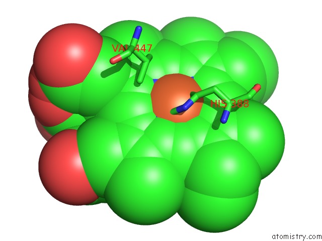
Mono view
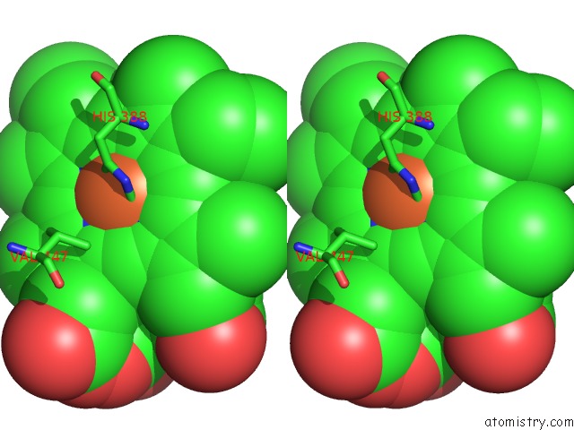
Stereo pair view
Mono view
Stereo pair view
A full contact list of Iron with other atoms in the Fe binding
site number 1 of 2.0 Angstrom Crystal Structure of Ovine Prostaglandin H2 Synthase-1, in Complex with Alpha-Methyl-4-Biphenylacetic Acid within 5.0Å range:
|
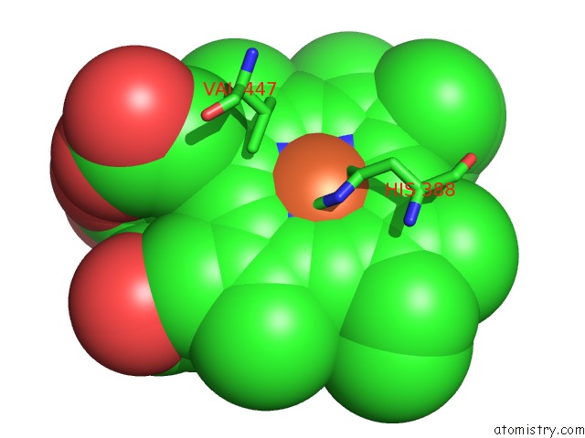
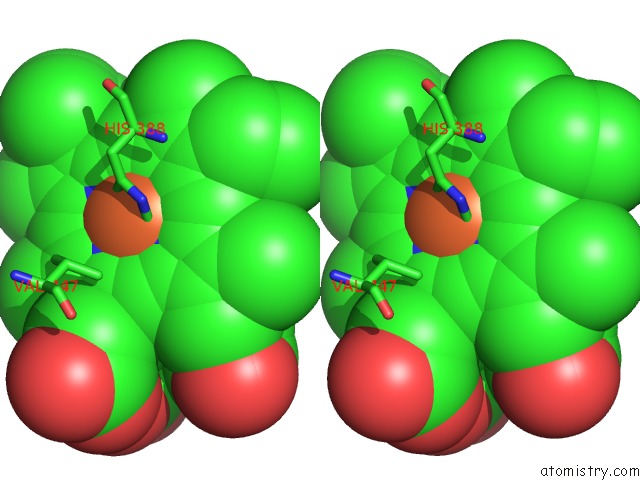
Iron binding site 2 out of 4 in 1q4g
Go back to
Iron binding site 2 out
of 4 in the 2.0 Angstrom Crystal Structure of Ovine Prostaglandin H2 Synthase-1, in Complex with Alpha-Methyl-4-Biphenylacetic Acid

Mono view

Stereo pair view
Mono view
Stereo pair view
A full contact list of Iron with other atoms in the Fe binding
site number 2 of 2.0 Angstrom Crystal Structure of Ovine Prostaglandin H2 Synthase-1, in Complex with Alpha-Methyl-4-Biphenylacetic Acid within 5.0Å range:
|
Iron binding site 3 out of 4 in 1q4g
Go back to
Iron binding site 3 out
of 4 in the 2.0 Angstrom Crystal Structure of Ovine Prostaglandin H2 Synthase-1, in Complex with Alpha-Methyl-4-Biphenylacetic Acid
Mono view
Stereo pair view
Mono view
Stereo pair view
A full contact list of Iron with other atoms in the Fe binding
site number 3 of 2.0 Angstrom Crystal Structure of Ovine Prostaglandin H2 Synthase-1, in Complex with Alpha-Methyl-4-Biphenylacetic Acid within 5.0Å range:
|
Iron binding site 4 out of 4 in 1q4g
Go back to
Iron binding site 4 out
of 4 in the 2.0 Angstrom Crystal Structure of Ovine Prostaglandin H2 Synthase-1, in Complex with Alpha-Methyl-4-Biphenylacetic Acid

Mono view

Stereo pair view
Mono view
Stereo pair view
A full contact list of Iron with other atoms in the Fe binding
site number 4 of 2.0 Angstrom Crystal Structure of Ovine Prostaglandin H2 Synthase-1, in Complex with Alpha-Methyl-4-Biphenylacetic Acid within 5.0Å range:
|
Reference:
K.Gupta,
B.S.Selinsky,
C.J.Kaub,
A.K.Katz,
P.J.Loll.
The 2.0A Resolution Crystal Structure of Prostaglandin H(2) Synthase-1: Structural Insights Into An Unusual Peroxidase J.Mol.Biol. V. 335 503 2004.
ISSN: ISSN 0022-2836
PubMed: 14672659
DOI: 10.1016/J.JMB.2003.10.073
Page generated: Wed Jul 16 19:44:39 2025
ISSN: ISSN 0022-2836
PubMed: 14672659
DOI: 10.1016/J.JMB.2003.10.073
Last articles
K in 8HZEK in 8HK6
K in 8HKK
K in 8HKF
K in 8HKM
K in 8HIR
K in 8GYR
K in 8GR9
K in 8H5L
K in 8H5J