Iron »
PDB 1q5e-1qom »
1q5e »
Iron in PDB 1q5e: Substrate-Free Cytochrome P450EPOK
Protein crystallography data
The structure of Substrate-Free Cytochrome P450EPOK, PDB code: 1q5e
was solved by
S.Nagano,
H.Li,
H.Shimizu,
C.Nishida,
H.Ogura,
P.R.Ortiz De Montellano,
T.L.Poulos,
with X-Ray Crystallography technique. A brief refinement statistics is given in the table below:
| Resolution Low / High (Å) | 50.00 / 2.65 |
| Space group | P 43 2 2 |
| Cell size a, b, c (Å), α, β, γ (°) | 61.520, 61.520, 256.730, 90.00, 90.00, 90.00 |
| R / Rfree (%) | 23.4 / 29.9 |
Iron Binding Sites:
The binding sites of Iron atom in the Substrate-Free Cytochrome P450EPOK
(pdb code 1q5e). This binding sites where shown within
5.0 Angstroms radius around Iron atom.
In total only one binding site of Iron was determined in the Substrate-Free Cytochrome P450EPOK, PDB code: 1q5e:
In total only one binding site of Iron was determined in the Substrate-Free Cytochrome P450EPOK, PDB code: 1q5e:
Iron binding site 1 out of 1 in 1q5e
Go back to
Iron binding site 1 out
of 1 in the Substrate-Free Cytochrome P450EPOK

Mono view
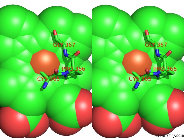
Stereo pair view
Mono view
Stereo pair view
A full contact list of Iron with other atoms in the Fe binding
site number 1 of Substrate-Free Cytochrome P450EPOK within 5.0Å range:
|
Reference:
S.Nagano,
H.Li,
H.Shimizu,
C.Nishida,
H.Ogura,
P.R.Ortiz De Montellano,
T.L.Poulos.
Crystal Structures of Epothilone D-Bound, Epothilone B-Bound, and Substrate-Free Forms of Cytochrome P450EPOK J.Biol.Chem. V. 278 44886 2003.
ISSN: ISSN 0021-9258
PubMed: 12933799
DOI: 10.1074/JBC.M308115200
Page generated: Wed Jul 16 19:47:12 2025
ISSN: ISSN 0021-9258
PubMed: 12933799
DOI: 10.1074/JBC.M308115200
Last articles
Mg in 4B5SMg in 4B4Z
Mg in 4B4X
Mg in 4B48
Mg in 4B47
Mg in 4B43
Mg in 4B3S
Mg in 4B2Q
Mg in 4B3A
Mg in 4B1Z