Iron »
PDB 1q5e-1qom »
1qf7 »
Iron in PDB 1qf7: Structure of the Mutant HIS392GLN of Catalase Hpii From E. Coli
Enzymatic activity of Structure of the Mutant HIS392GLN of Catalase Hpii From E. Coli
All present enzymatic activity of Structure of the Mutant HIS392GLN of Catalase Hpii From E. Coli:
1.11.1.6;
1.11.1.6;
Protein crystallography data
The structure of Structure of the Mutant HIS392GLN of Catalase Hpii From E. Coli, PDB code: 1qf7
was solved by
M.J.Mate,
P.C.Loewen,
I.Fita,
with X-Ray Crystallography technique. A brief refinement statistics is given in the table below:
| Resolution Low / High (Å) | 20.00 / 2.20 |
| Space group | P 1 21 1 |
| Cell size a, b, c (Å), α, β, γ (°) | 93.400, 132.920, 121.670, 90.00, 109.47, 90.00 |
| R / Rfree (%) | 14.4 / 21 |
Iron Binding Sites:
The binding sites of Iron atom in the Structure of the Mutant HIS392GLN of Catalase Hpii From E. Coli
(pdb code 1qf7). This binding sites where shown within
5.0 Angstroms radius around Iron atom.
In total 4 binding sites of Iron where determined in the Structure of the Mutant HIS392GLN of Catalase Hpii From E. Coli, PDB code: 1qf7:
Jump to Iron binding site number: 1; 2; 3; 4;
In total 4 binding sites of Iron where determined in the Structure of the Mutant HIS392GLN of Catalase Hpii From E. Coli, PDB code: 1qf7:
Jump to Iron binding site number: 1; 2; 3; 4;
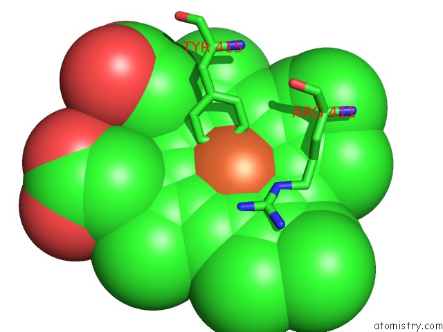
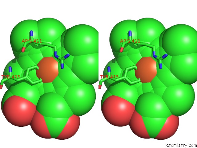
Iron binding site 1 out of 4 in 1qf7
Go back to
Iron binding site 1 out
of 4 in the Structure of the Mutant HIS392GLN of Catalase Hpii From E. Coli
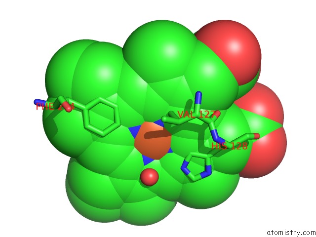
Mono view
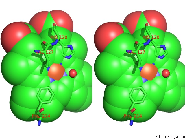
Stereo pair view
Mono view
Stereo pair view
A full contact list of Iron with other atoms in the Fe binding
site number 1 of Structure of the Mutant HIS392GLN of Catalase Hpii From E. Coli within 5.0Å range:
|
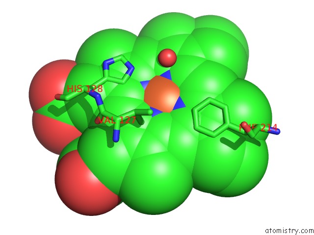
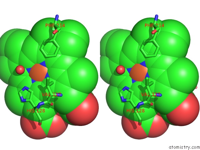
Iron binding site 2 out of 4 in 1qf7
Go back to
Iron binding site 2 out
of 4 in the Structure of the Mutant HIS392GLN of Catalase Hpii From E. Coli
Mono view
Stereo pair view
Mono view
Stereo pair view
A full contact list of Iron with other atoms in the Fe binding
site number 2 of Structure of the Mutant HIS392GLN of Catalase Hpii From E. Coli within 5.0Å range:
|
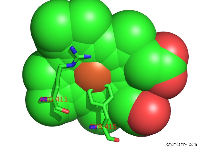
Iron binding site 3 out of 4 in 1qf7
Go back to
Iron binding site 3 out
of 4 in the Structure of the Mutant HIS392GLN of Catalase Hpii From E. Coli
Mono view
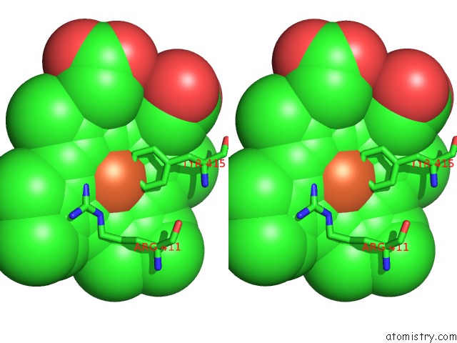
Stereo pair view
Mono view
Stereo pair view
A full contact list of Iron with other atoms in the Fe binding
site number 3 of Structure of the Mutant HIS392GLN of Catalase Hpii From E. Coli within 5.0Å range:
|
Iron binding site 4 out of 4 in 1qf7
Go back to
Iron binding site 4 out
of 4 in the Structure of the Mutant HIS392GLN of Catalase Hpii From E. Coli
Mono view
Stereo pair view
Mono view
Stereo pair view
A full contact list of Iron with other atoms in the Fe binding
site number 4 of Structure of the Mutant HIS392GLN of Catalase Hpii From E. Coli within 5.0Å range:
|
Reference:
M.J.Mate,
M.S.Sevinc,
B.Hu,
J.Bujons,
J.Bravo,
J.Switala,
W.Ens,
P.C.Loewen,
I.Fita.
Mutants That Alter the Covalent Structure of Catalase Hydroperoxidase II From Escherichia Coli. J.Biol.Chem. V. 274 27717 1999.
ISSN: ISSN 0021-9258
PubMed: 10488114
DOI: 10.1074/JBC.274.39.27717
Page generated: Wed Jul 16 19:47:37 2025
ISSN: ISSN 0021-9258
PubMed: 10488114
DOI: 10.1074/JBC.274.39.27717
Last articles
Mg in 3D19Mg in 3CZ4
Mg in 3CZ1
Mg in 3CZ0
Mg in 3CXO
Mg in 3CYZ
Mg in 3CYI
Mg in 3CX8
Mg in 3CX7
Mg in 3CX6