Iron »
PDB 1q5e-1qom »
1qi8 »
Iron in PDB 1qi8: Deoxygenated Structure of A Distal Pocket Hemoglobin Mutant
Protein crystallography data
The structure of Deoxygenated Structure of A Distal Pocket Hemoglobin Mutant, PDB code: 1qi8
was solved by
A.E.Miele,
B.Vallone,
S.Santanche,
C.Travaglini-Allocatelli,
A.Bellelli,
M.Brunori,
with X-Ray Crystallography technique. A brief refinement statistics is given in the table below:
| Resolution Low / High (Å) | 16.00 / 1.80 |
| Space group | P 1 21 1 |
| Cell size a, b, c (Å), α, β, γ (°) | 63.360, 94.320, 54.000, 90.00, 99.43, 90.00 |
| R / Rfree (%) | 16.9 / 22.2 |
Iron Binding Sites:
The binding sites of Iron atom in the Deoxygenated Structure of A Distal Pocket Hemoglobin Mutant
(pdb code 1qi8). This binding sites where shown within
5.0 Angstroms radius around Iron atom.
In total 4 binding sites of Iron where determined in the Deoxygenated Structure of A Distal Pocket Hemoglobin Mutant, PDB code: 1qi8:
Jump to Iron binding site number: 1; 2; 3; 4;
In total 4 binding sites of Iron where determined in the Deoxygenated Structure of A Distal Pocket Hemoglobin Mutant, PDB code: 1qi8:
Jump to Iron binding site number: 1; 2; 3; 4;
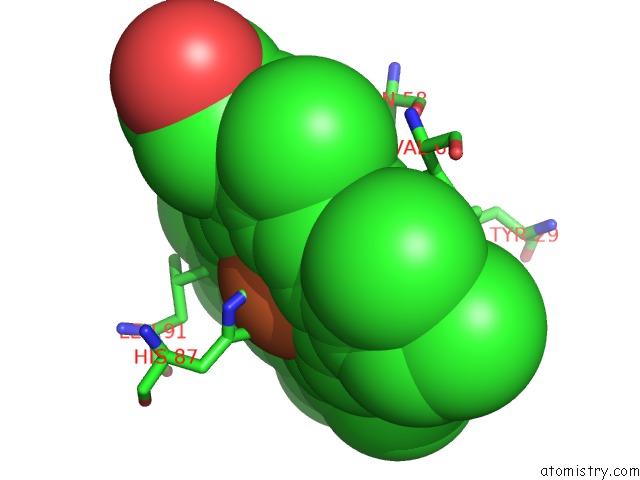
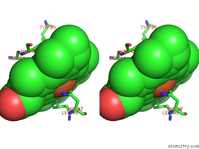
Iron binding site 1 out of 4 in 1qi8
Go back to
Iron binding site 1 out
of 4 in the Deoxygenated Structure of A Distal Pocket Hemoglobin Mutant
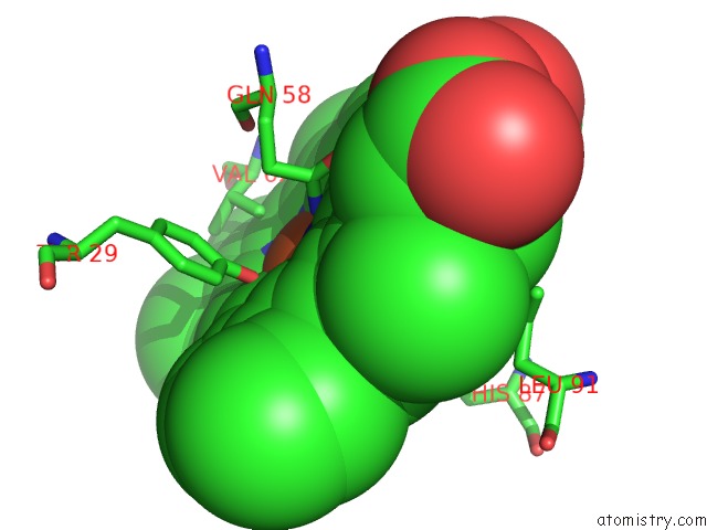
Mono view
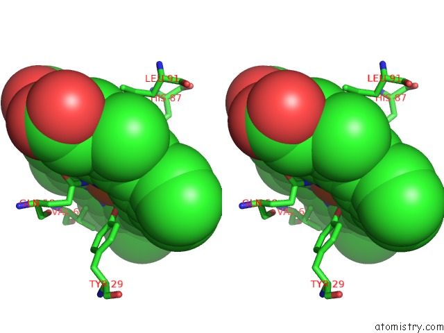
Stereo pair view
Mono view
Stereo pair view
A full contact list of Iron with other atoms in the Fe binding
site number 1 of Deoxygenated Structure of A Distal Pocket Hemoglobin Mutant within 5.0Å range:
|
Iron binding site 2 out of 4 in 1qi8
Go back to
Iron binding site 2 out
of 4 in the Deoxygenated Structure of A Distal Pocket Hemoglobin Mutant
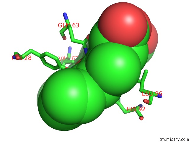
Mono view
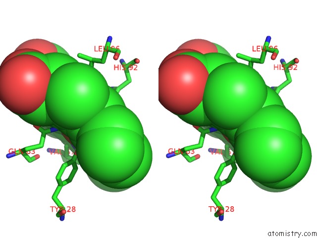
Stereo pair view
Mono view
Stereo pair view
A full contact list of Iron with other atoms in the Fe binding
site number 2 of Deoxygenated Structure of A Distal Pocket Hemoglobin Mutant within 5.0Å range:
|
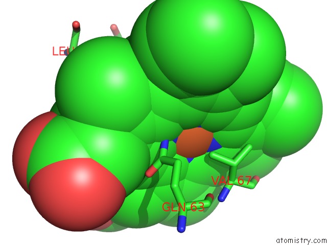
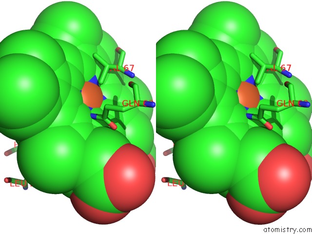
Iron binding site 3 out of 4 in 1qi8
Go back to
Iron binding site 3 out
of 4 in the Deoxygenated Structure of A Distal Pocket Hemoglobin Mutant
Mono view
Stereo pair view
Mono view
Stereo pair view
A full contact list of Iron with other atoms in the Fe binding
site number 3 of Deoxygenated Structure of A Distal Pocket Hemoglobin Mutant within 5.0Å range:
|
Iron binding site 4 out of 4 in 1qi8
Go back to
Iron binding site 4 out
of 4 in the Deoxygenated Structure of A Distal Pocket Hemoglobin Mutant
Mono view
Stereo pair view
Mono view
Stereo pair view
A full contact list of Iron with other atoms in the Fe binding
site number 4 of Deoxygenated Structure of A Distal Pocket Hemoglobin Mutant within 5.0Å range:
|
Reference:
A.E.Miele,
S.Santanche,
C.Travaglini-Allocatelli,
B.Vallone,
M.Brunori,
A.Bellelli.
Modulation of Ligand Binding in Engineered Human Hemoglobin Distal Pocket. J.Mol.Biol. V. 290 515 1999.
ISSN: ISSN 0022-2836
PubMed: 10390349
DOI: 10.1006/JMBI.1999.2869
Page generated: Wed Jul 16 19:52:55 2025
ISSN: ISSN 0022-2836
PubMed: 10390349
DOI: 10.1006/JMBI.1999.2869
Last articles
Mg in 4Z4GMg in 4Z4E
Mg in 4Z4F
Mg in 4Z4D
Mg in 4Z40
Mg in 4Z4C
Mg in 4Z3Z
Mg in 4Z4B
Mg in 4Z3Y
Mg in 4Z3X