Iron »
PDB 1rak-1rsv »
1raq »
Iron in PDB 1raq: The Structure and Function of Omega Loop A Replacements in Cytochrome C
Protein crystallography data
The structure of The Structure and Function of Omega Loop A Replacements in Cytochrome C, PDB code: 1raq
was solved by
M.E.P.Murphy,
G.D.Brayer,
with X-Ray Crystallography technique. A brief refinement statistics is given in the table below:
| Resolution Low / High (Å) | N/A / 1.90 |
| Space group | P 43 21 2 |
| Cell size a, b, c (Å), α, β, γ (°) | 36.370, 36.370, 137.640, 90.00, 90.00, 90.00 |
| R / Rfree (%) | n/a / n/a |
Iron Binding Sites:
The binding sites of Iron atom in the The Structure and Function of Omega Loop A Replacements in Cytochrome C
(pdb code 1raq). This binding sites where shown within
5.0 Angstroms radius around Iron atom.
In total only one binding site of Iron was determined in the The Structure and Function of Omega Loop A Replacements in Cytochrome C, PDB code: 1raq:
In total only one binding site of Iron was determined in the The Structure and Function of Omega Loop A Replacements in Cytochrome C, PDB code: 1raq:
Iron binding site 1 out of 1 in 1raq
Go back to
Iron binding site 1 out
of 1 in the The Structure and Function of Omega Loop A Replacements in Cytochrome C
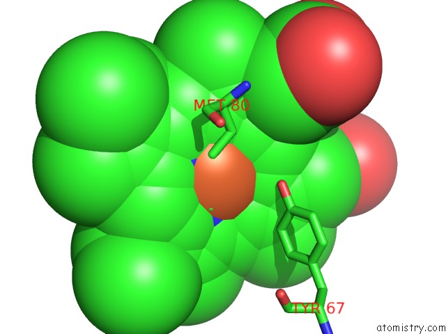
Mono view
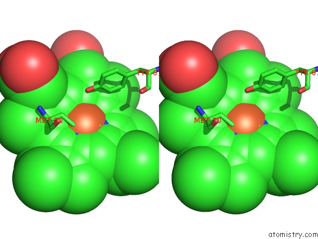
Stereo pair view
Mono view
Stereo pair view
A full contact list of Iron with other atoms in the Fe binding
site number 1 of The Structure and Function of Omega Loop A Replacements in Cytochrome C within 5.0Å range:
|
Reference:
M.E.Murphy,
J.S.Fetrow,
R.E.Burton,
G.D.Brayer.
The Structure and Function of Omega Loop A Replacements in Cytochrome C. Protein Sci. V. 2 1429 1993.
ISSN: ISSN 0961-8368
PubMed: 8401228
Page generated: Wed Jul 16 20:20:59 2025
ISSN: ISSN 0961-8368
PubMed: 8401228
Last articles
Mg in 5ORPMg in 5ORO
Mg in 5ORN
Mg in 5ORL
Mg in 5ORK
Mg in 5OPX
Mg in 5ORJ
Mg in 5OQU
Mg in 5OQM
Mg in 5OQJ