Iron »
PDB 1rak-1rsv »
1rgv »
Iron in PDB 1rgv: Crystal Structure of the Ferredoxin From Thauera Aromatica
Protein crystallography data
The structure of Crystal Structure of the Ferredoxin From Thauera Aromatica, PDB code: 1rgv
was solved by
M.Unciuleac,
M.Boll,
E.Warkentin,
U.Ermler,
with X-Ray Crystallography technique. A brief refinement statistics is given in the table below:
| Resolution Low / High (Å) | 25.86 / 2.90 |
| Space group | P 31 2 1 |
| Cell size a, b, c (Å), α, β, γ (°) | 79.000, 79.000, 49.300, 90.00, 90.00, 120.00 |
| R / Rfree (%) | 19.7 / 21.9 |
Iron Binding Sites:
The binding sites of Iron atom in the Crystal Structure of the Ferredoxin From Thauera Aromatica
(pdb code 1rgv). This binding sites where shown within
5.0 Angstroms radius around Iron atom.
In total 8 binding sites of Iron where determined in the Crystal Structure of the Ferredoxin From Thauera Aromatica, PDB code: 1rgv:
Jump to Iron binding site number: 1; 2; 3; 4; 5; 6; 7; 8;
In total 8 binding sites of Iron where determined in the Crystal Structure of the Ferredoxin From Thauera Aromatica, PDB code: 1rgv:
Jump to Iron binding site number: 1; 2; 3; 4; 5; 6; 7; 8;
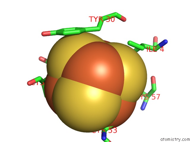
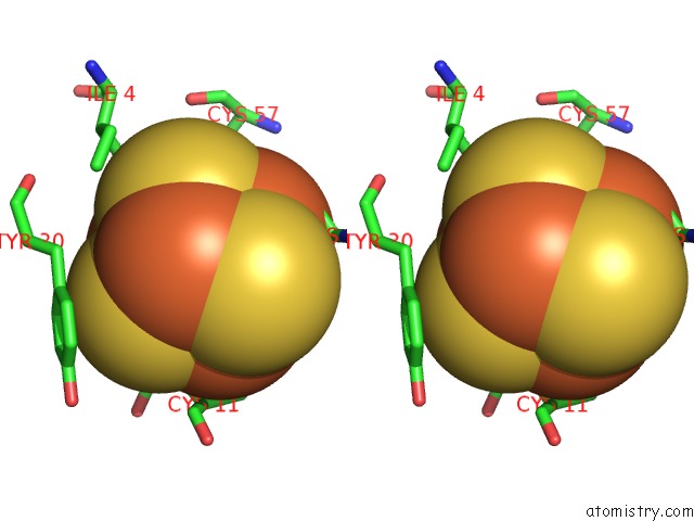
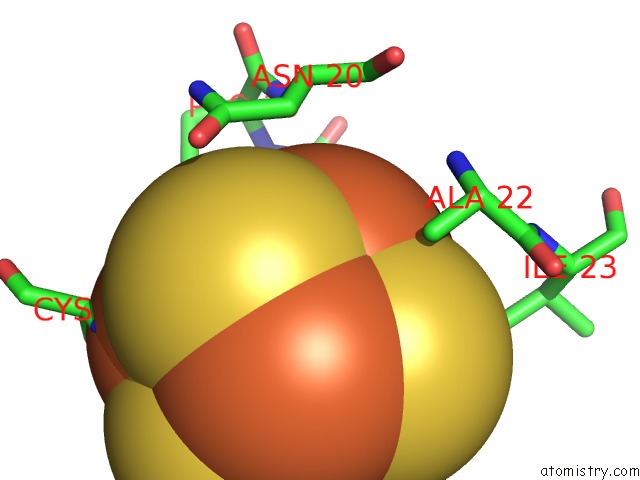
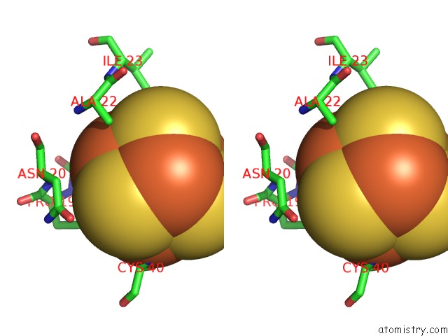
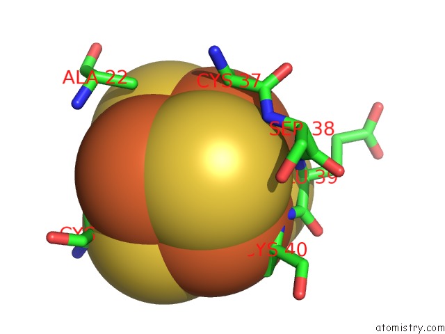
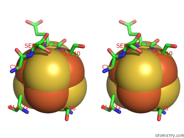
Iron binding site 1 out of 8 in 1rgv
Go back to
Iron binding site 1 out
of 8 in the Crystal Structure of the Ferredoxin From Thauera Aromatica
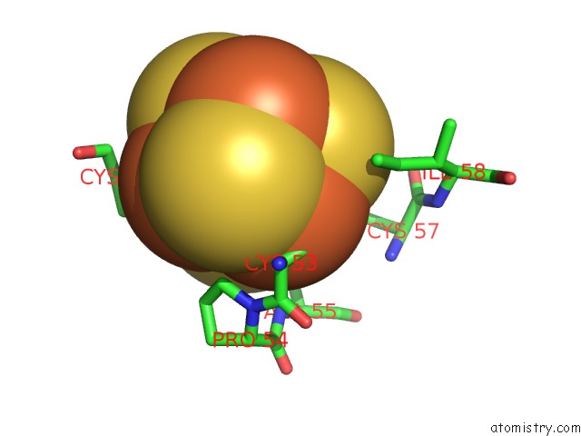
Mono view
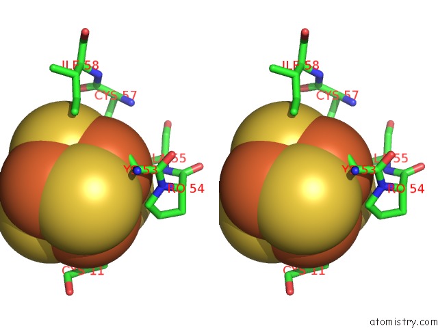
Stereo pair view
Mono view
Stereo pair view
A full contact list of Iron with other atoms in the Fe binding
site number 1 of Crystal Structure of the Ferredoxin From Thauera Aromatica within 5.0Å range:
|
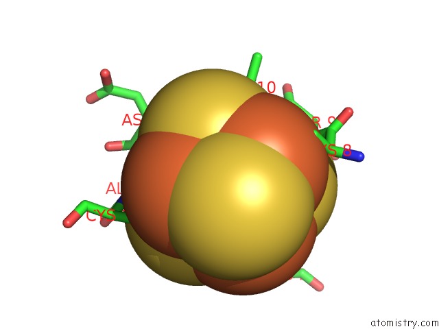
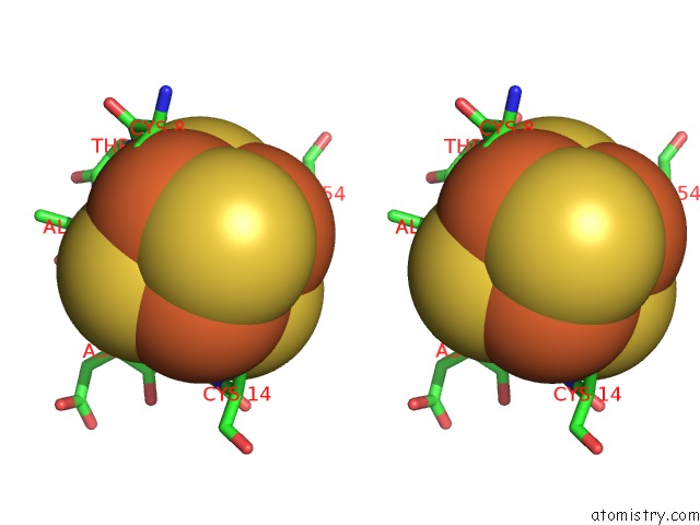
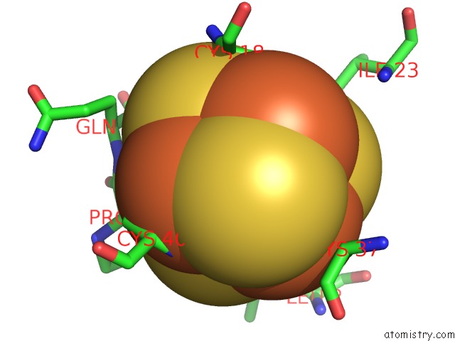
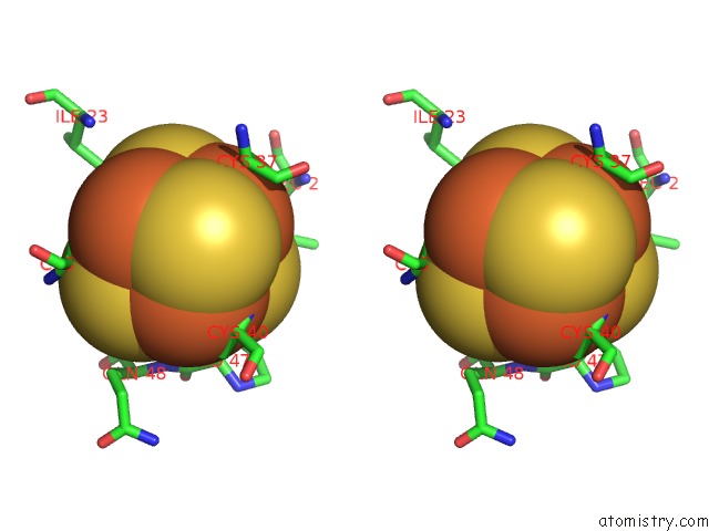
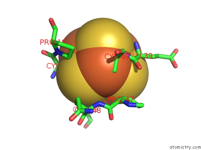
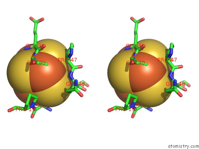
Iron binding site 2 out of 8 in 1rgv
Go back to
Iron binding site 2 out
of 8 in the Crystal Structure of the Ferredoxin From Thauera Aromatica
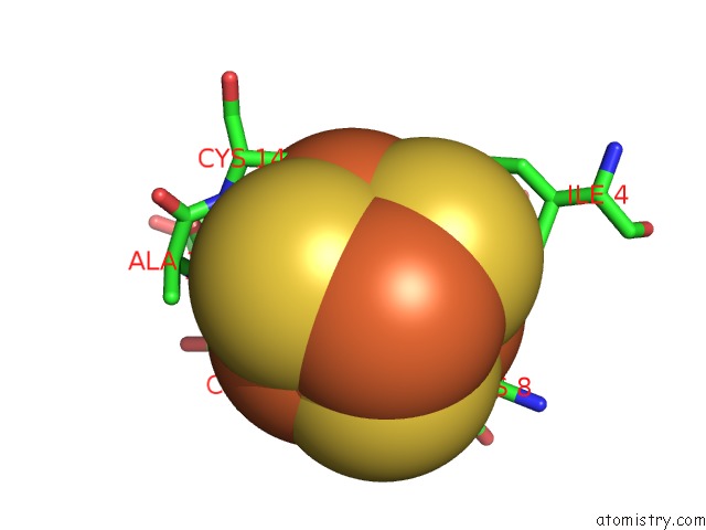
Mono view
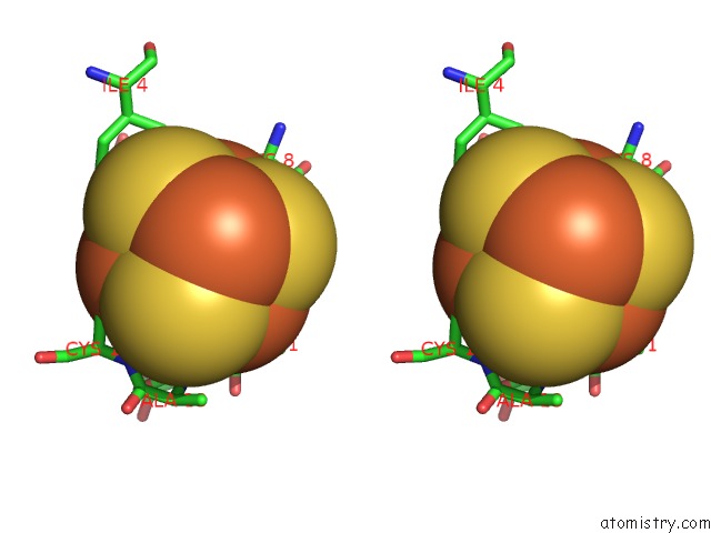
Stereo pair view
Mono view
Stereo pair view
A full contact list of Iron with other atoms in the Fe binding
site number 2 of Crystal Structure of the Ferredoxin From Thauera Aromatica within 5.0Å range:
|
Iron binding site 3 out of 8 in 1rgv
Go back to
Iron binding site 3 out
of 8 in the Crystal Structure of the Ferredoxin From Thauera Aromatica
Mono view
Stereo pair view
Mono view
Stereo pair view
A full contact list of Iron with other atoms in the Fe binding
site number 3 of Crystal Structure of the Ferredoxin From Thauera Aromatica within 5.0Å range:
|
Iron binding site 4 out of 8 in 1rgv
Go back to
Iron binding site 4 out
of 8 in the Crystal Structure of the Ferredoxin From Thauera Aromatica
Mono view
Stereo pair view
Mono view
Stereo pair view
A full contact list of Iron with other atoms in the Fe binding
site number 4 of Crystal Structure of the Ferredoxin From Thauera Aromatica within 5.0Å range:
|
Iron binding site 5 out of 8 in 1rgv
Go back to
Iron binding site 5 out
of 8 in the Crystal Structure of the Ferredoxin From Thauera Aromatica
Mono view
Stereo pair view
Mono view
Stereo pair view
A full contact list of Iron with other atoms in the Fe binding
site number 5 of Crystal Structure of the Ferredoxin From Thauera Aromatica within 5.0Å range:
|
Iron binding site 6 out of 8 in 1rgv
Go back to
Iron binding site 6 out
of 8 in the Crystal Structure of the Ferredoxin From Thauera Aromatica
Mono view
Stereo pair view
Mono view
Stereo pair view
A full contact list of Iron with other atoms in the Fe binding
site number 6 of Crystal Structure of the Ferredoxin From Thauera Aromatica within 5.0Å range:
|
Iron binding site 7 out of 8 in 1rgv
Go back to
Iron binding site 7 out
of 8 in the Crystal Structure of the Ferredoxin From Thauera Aromatica
Mono view
Stereo pair view
Mono view
Stereo pair view
A full contact list of Iron with other atoms in the Fe binding
site number 7 of Crystal Structure of the Ferredoxin From Thauera Aromatica within 5.0Å range:
|
Iron binding site 8 out of 8 in 1rgv
Go back to
Iron binding site 8 out
of 8 in the Crystal Structure of the Ferredoxin From Thauera Aromatica
Mono view
Stereo pair view
Mono view
Stereo pair view
A full contact list of Iron with other atoms in the Fe binding
site number 8 of Crystal Structure of the Ferredoxin From Thauera Aromatica within 5.0Å range:
|
Reference:
M.Unciuleac,
M.Boll,
E.Warkentin,
U.Ermler.
Crystallization of 4-Hydroxybenzoyl-Coa Reductase and the Structure of Its Electron Donor Ferredoxin. Acta Crystallogr.,Sect.D V. 60 388 2004.
ISSN: ISSN 0907-4449
PubMed: 14747735
DOI: 10.1107/S0907444903028506
Page generated: Wed Jul 16 20:22:39 2025
ISSN: ISSN 0907-4449
PubMed: 14747735
DOI: 10.1107/S0907444903028506
Last articles
Mg in 5PJUMg in 5PJW
Mg in 5PJV
Mg in 5PJT
Mg in 5PJQ
Mg in 5PJS
Mg in 5PJP
Mg in 5PJR
Mg in 5PJL
Mg in 5PJN