Iron »
PDB 1rte-1sdk »
1rxf »
Iron in PDB 1rxf: Deacetoxycephalosporin C Synthase Complexed with Fe(II)
Protein crystallography data
The structure of Deacetoxycephalosporin C Synthase Complexed with Fe(II), PDB code: 1rxf
was solved by
K.Valegard,
A.C.Terwisscha Van Scheltinga,
M.D.Lloyd,
T.Hara,
S.Ramaswamy,
A.Perrakis,
A.Thompson,
H.J.Lee,
J.E.Baldwin,
C.J.Schofield,
J.Hajdu,
I.Andersson,
with X-Ray Crystallography technique. A brief refinement statistics is given in the table below:
| Resolution Low / High (Å) | 20.00 / 1.50 |
| Space group | H 3 |
| Cell size a, b, c (Å), α, β, γ (°) | 106.100, 106.100, 71.200, 90.00, 90.00, 120.00 |
| R / Rfree (%) | 11.9 / 14.2 |
Iron Binding Sites:
The binding sites of Iron atom in the Deacetoxycephalosporin C Synthase Complexed with Fe(II)
(pdb code 1rxf). This binding sites where shown within
5.0 Angstroms radius around Iron atom.
In total only one binding site of Iron was determined in the Deacetoxycephalosporin C Synthase Complexed with Fe(II), PDB code: 1rxf:
In total only one binding site of Iron was determined in the Deacetoxycephalosporin C Synthase Complexed with Fe(II), PDB code: 1rxf:
Iron binding site 1 out of 1 in 1rxf
Go back to
Iron binding site 1 out
of 1 in the Deacetoxycephalosporin C Synthase Complexed with Fe(II)
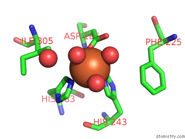
Mono view
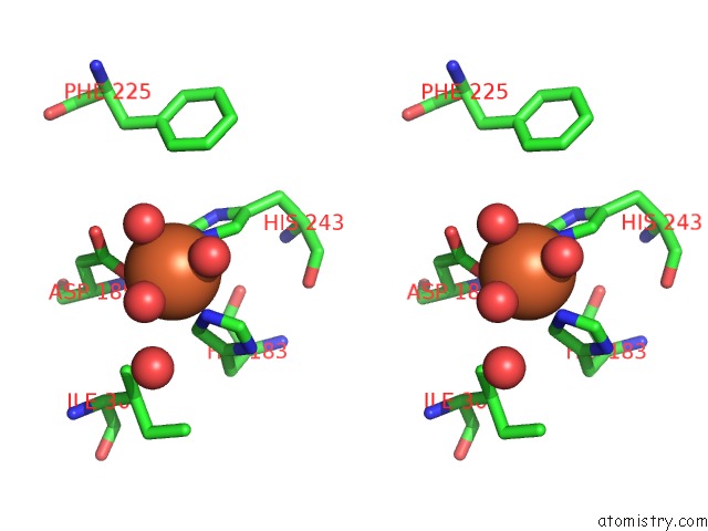
Stereo pair view
Mono view
Stereo pair view
A full contact list of Iron with other atoms in the Fe binding
site number 1 of Deacetoxycephalosporin C Synthase Complexed with Fe(II) within 5.0Å range:
|
Reference:
K.Valegard,
A.C.Van Scheltinga,
M.D.Lloyd,
T.Hara,
S.Ramaswamy,
A.Perrakis,
A.Thompson,
H.J.Lee,
J.E.Baldwin,
C.J.Schofield,
J.Hajdu,
I.Andersson.
Structure of A Cephalosporin Synthase. Nature V. 394 805 1998.
ISSN: ISSN 0028-0836
PubMed: 9723623
DOI: 10.1038/29575
Page generated: Wed Jul 16 20:31:35 2025
ISSN: ISSN 0028-0836
PubMed: 9723623
DOI: 10.1038/29575
Last articles
Mn in 9LJUMn in 9LJW
Mn in 9LJS
Mn in 9LJR
Mn in 9LJT
Mn in 9LJV
Mg in 9UA2
Mg in 9R96
Mg in 9VM1
Mg in 9P01