Iron »
PDB 1rte-1sdk »
1ryt »
Iron in PDB 1ryt: Rubrerythrin
Protein crystallography data
The structure of Rubrerythrin, PDB code: 1ryt
was solved by
F.Demare,
D.M.Kurtz,
P.Nordlund,
with X-Ray Crystallography technique. A brief refinement statistics is given in the table below:
| Resolution Low / High (Å) | 8.00 / 2.10 |
| Space group | I 2 2 2 |
| Cell size a, b, c (Å), α, β, γ (°) | 50.400, 82.000, 100.700, 90.00, 90.00, 90.00 |
| R / Rfree (%) | 18.2 / 25.6 |
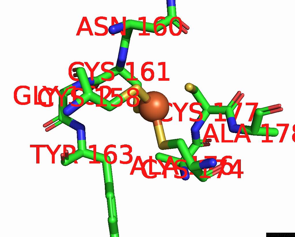
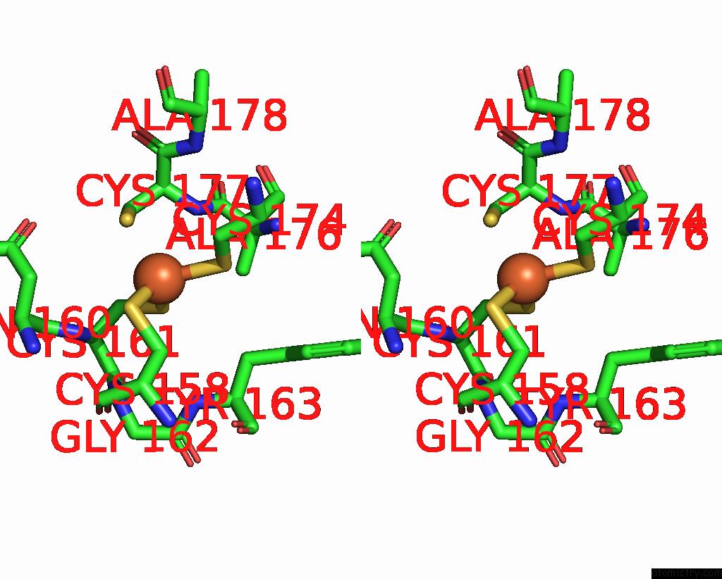
Iron Binding Sites:
The binding sites of Iron atom in the Rubrerythrin
(pdb code 1ryt). This binding sites where shown within
5.0 Angstroms radius around Iron atom.
In total 3 binding sites of Iron where determined in the Rubrerythrin, PDB code: 1ryt:
Jump to Iron binding site number: 1; 2; 3;
In total 3 binding sites of Iron where determined in the Rubrerythrin, PDB code: 1ryt:
Jump to Iron binding site number: 1; 2; 3;
Iron binding site 1 out of 3 in 1ryt
Go back to
Iron binding site 1 out
of 3 in the Rubrerythrin
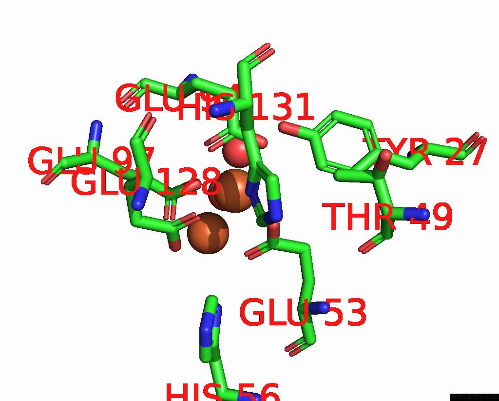
Mono view
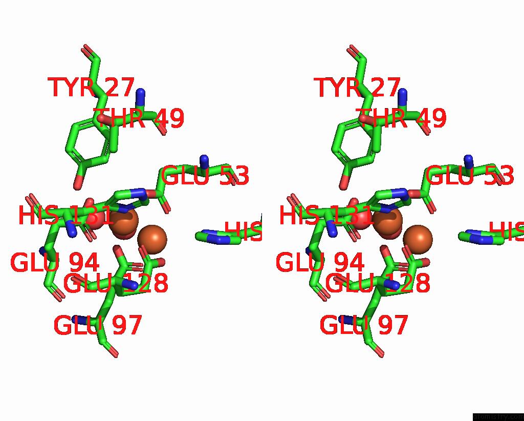
Stereo pair view
Mono view
Stereo pair view
A full contact list of Iron with other atoms in the Fe binding
site number 1 of Rubrerythrin within 5.0Å range:
|
Iron binding site 2 out of 3 in 1ryt
Go back to
Iron binding site 2 out
of 3 in the Rubrerythrin
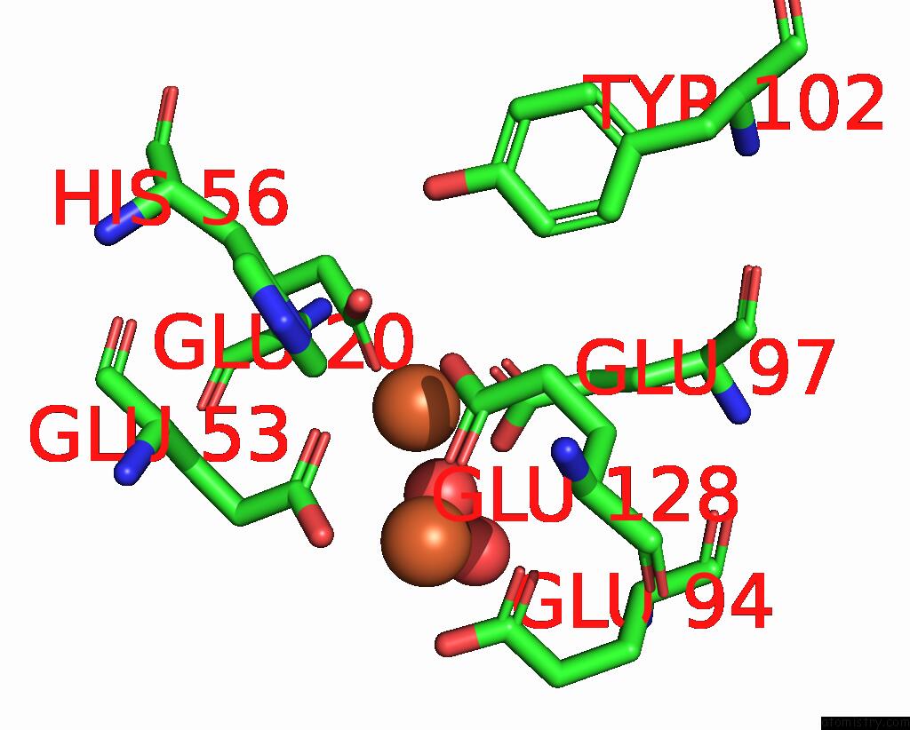
Mono view
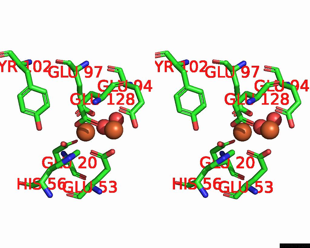
Stereo pair view
Mono view
Stereo pair view
A full contact list of Iron with other atoms in the Fe binding
site number 2 of Rubrerythrin within 5.0Å range:
|
Iron binding site 3 out of 3 in 1ryt
Go back to
Iron binding site 3 out
of 3 in the Rubrerythrin
Mono view
Stereo pair view
Mono view
Stereo pair view
A full contact list of Iron with other atoms in the Fe binding
site number 3 of Rubrerythrin within 5.0Å range:
|
Reference:
F.Demare,
D.M.Kurtz Jr.,
P.Nordlund.
The Structure of Desulfovibrio Vulgaris Rubrerythrin Reveals A Unique Combination of Rubredoxin-Like FES4 and Ferritin-Like Diiron Domains. Nat.Struct.Biol. V. 3 539 1996.
ISSN: ISSN 1072-8368
PubMed: 8646540
DOI: 10.1038/NSB0696-539
Page generated: Wed Jul 16 20:32:07 2025
ISSN: ISSN 1072-8368
PubMed: 8646540
DOI: 10.1038/NSB0696-539
Last articles
Mn in 9LJUMn in 9LJW
Mn in 9LJS
Mn in 9LJR
Mn in 9LJT
Mn in 9LJV
Mg in 9UA2
Mg in 9R96
Mg in 9VM1
Mg in 9P01