Iron »
PDB 1rte-1sdk »
1s6a »
Iron in PDB 1s6a: The X-Ray Structure of the Cyanobacteria Synechocystis Hemoglobin "Cyanoglobin" with Azide Ligand
Protein crystallography data
The structure of The X-Ray Structure of the Cyanobacteria Synechocystis Hemoglobin "Cyanoglobin" with Azide Ligand, PDB code: 1s6a
was solved by
J.T.Trent Iii,
S.Kundu,
J.A.Hoy,
M.S.Hargrove,
with X-Ray Crystallography technique. A brief refinement statistics is given in the table below:
| Resolution Low / High (Å) | 32.40 / 1.69 |
| Space group | I 41 |
| Cell size a, b, c (Å), α, β, γ (°) | 83.350, 83.350, 65.180, 90.00, 90.00, 90.00 |
| R / Rfree (%) | 19.7 / 21.9 |
Iron Binding Sites:
The binding sites of Iron atom in the The X-Ray Structure of the Cyanobacteria Synechocystis Hemoglobin "Cyanoglobin" with Azide Ligand
(pdb code 1s6a). This binding sites where shown within
5.0 Angstroms radius around Iron atom.
In total only one binding site of Iron was determined in the The X-Ray Structure of the Cyanobacteria Synechocystis Hemoglobin "Cyanoglobin" with Azide Ligand, PDB code: 1s6a:
In total only one binding site of Iron was determined in the The X-Ray Structure of the Cyanobacteria Synechocystis Hemoglobin "Cyanoglobin" with Azide Ligand, PDB code: 1s6a:
Iron binding site 1 out of 1 in 1s6a
Go back to
Iron binding site 1 out
of 1 in the The X-Ray Structure of the Cyanobacteria Synechocystis Hemoglobin "Cyanoglobin" with Azide Ligand
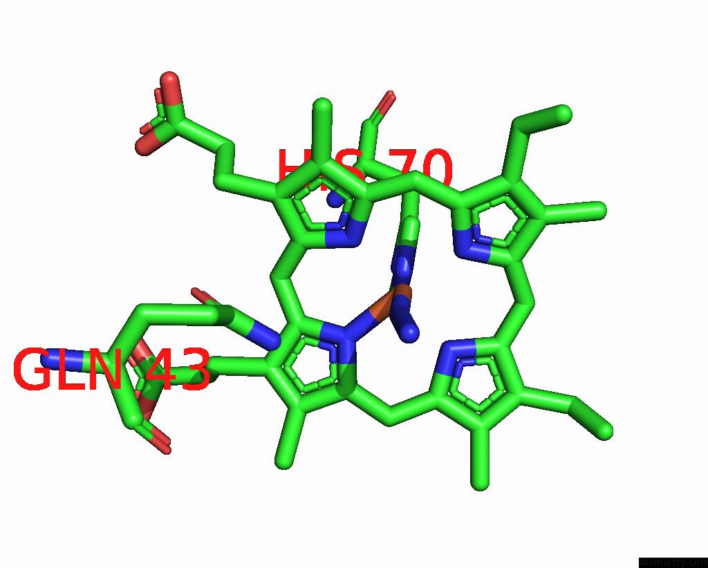
Mono view
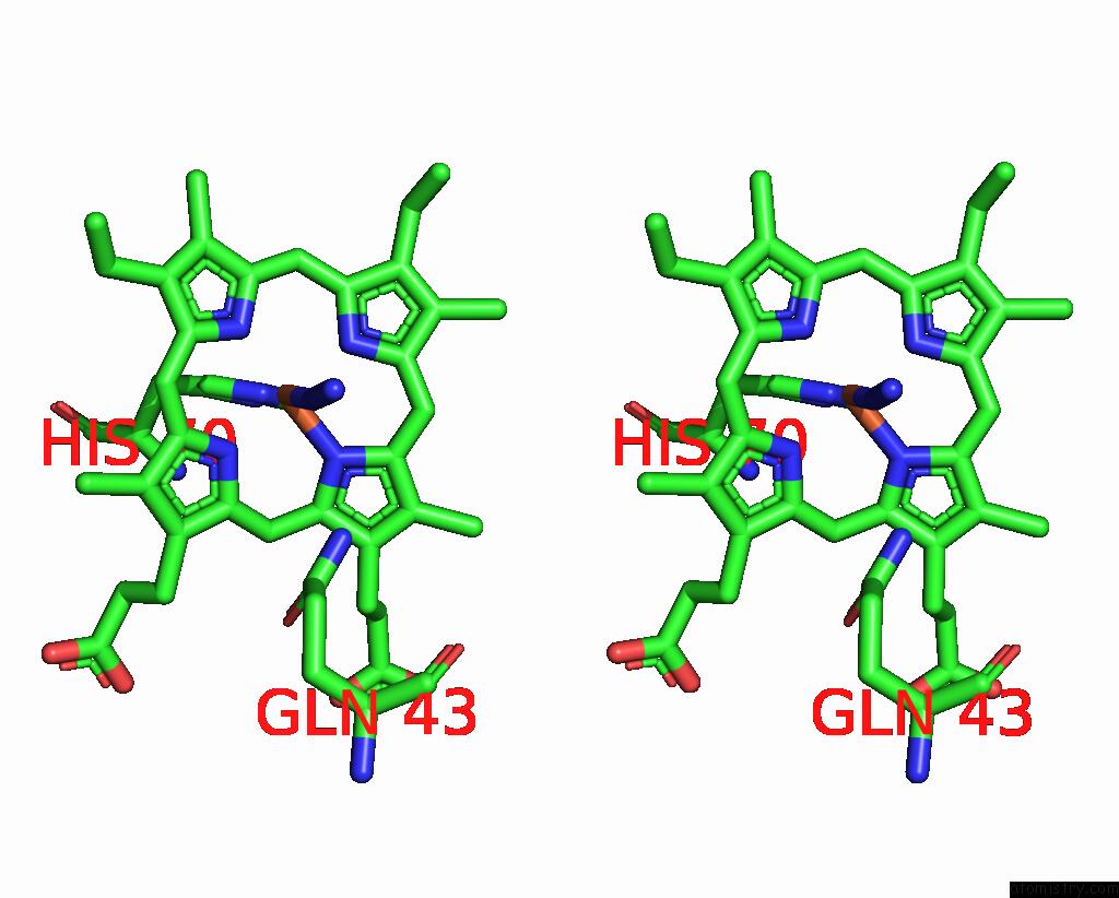
Stereo pair view
Mono view
Stereo pair view
A full contact list of Iron with other atoms in the Fe binding
site number 1 of The X-Ray Structure of the Cyanobacteria Synechocystis Hemoglobin "Cyanoglobin" with Azide Ligand within 5.0Å range:
|
Reference:
J.T.Trent Iii,
S.Kundu,
J.A.Hoy,
M.S.Hargrove.
Crystallographic Analysis of Synechocystis Cyanoglobin Reveals the Structural Changes Accompanying Ligand Binding in A Hexacoordinate Hemoglobin. J.Mol.Biol. V. 341 1097 2004.
ISSN: ISSN 0022-2836
PubMed: 15289104
DOI: 10.1016/J.JMB.2004.05.070
Page generated: Wed Jul 16 20:35:17 2025
ISSN: ISSN 0022-2836
PubMed: 15289104
DOI: 10.1016/J.JMB.2004.05.070
Last articles
Mn in 9LJUMn in 9LJW
Mn in 9LJS
Mn in 9LJR
Mn in 9LJT
Mn in 9LJV
Mg in 9UA2
Mg in 9R96
Mg in 9VM1
Mg in 9P01