Iron »
PDB 1rte-1sdk »
1sch »
Iron in PDB 1sch: Peanut Peroxidase
Enzymatic activity of Peanut Peroxidase
All present enzymatic activity of Peanut Peroxidase:
1.11.1.7;
1.11.1.7;
Protein crystallography data
The structure of Peanut Peroxidase, PDB code: 1sch
was solved by
D.J.Schuller,
T.L.Poulos,
with X-Ray Crystallography technique. A brief refinement statistics is given in the table below:
| Resolution Low / High (Å) | 10.00 / 2.56 |
| Space group | P 21 21 21 |
| Cell size a, b, c (Å), α, β, γ (°) | 48.100, 97.200, 146.200, 90.00, 90.00, 90.00 |
| R / Rfree (%) | 19.9 / 26.2 |
Other elements in 1sch:
The structure of Peanut Peroxidase also contains other interesting chemical elements:
| Calcium | (Ca) | 4 atoms |
Iron Binding Sites:
The binding sites of Iron atom in the Peanut Peroxidase
(pdb code 1sch). This binding sites where shown within
5.0 Angstroms radius around Iron atom.
In total 2 binding sites of Iron where determined in the Peanut Peroxidase, PDB code: 1sch:
Jump to Iron binding site number: 1; 2;
In total 2 binding sites of Iron where determined in the Peanut Peroxidase, PDB code: 1sch:
Jump to Iron binding site number: 1; 2;
Iron binding site 1 out of 2 in 1sch
Go back to
Iron binding site 1 out
of 2 in the Peanut Peroxidase
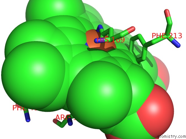
Mono view
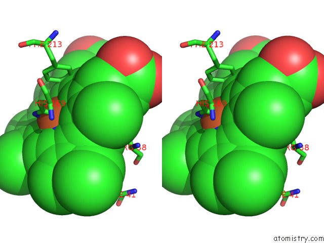
Stereo pair view
Mono view
Stereo pair view
A full contact list of Iron with other atoms in the Fe binding
site number 1 of Peanut Peroxidase within 5.0Å range:
|
Iron binding site 2 out of 2 in 1sch
Go back to
Iron binding site 2 out
of 2 in the Peanut Peroxidase
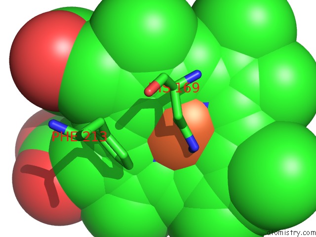
Mono view
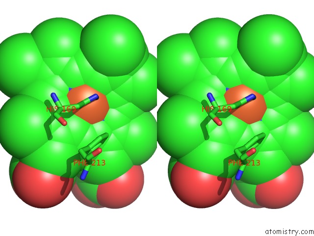
Stereo pair view
Mono view
Stereo pair view
A full contact list of Iron with other atoms in the Fe binding
site number 2 of Peanut Peroxidase within 5.0Å range:
|
Reference:
D.J.Schuller,
N.Ban,
R.B.Huystee,
A.Mcpherson,
T.L.Poulos.
The Crystal Structure of Peanut Peroxidase. Structure V. 4 311 1996.
ISSN: ISSN 0969-2126
PubMed: 8805539
DOI: 10.1016/S0969-2126(96)00035-4
Page generated: Wed Jul 16 20:36:02 2025
ISSN: ISSN 0969-2126
PubMed: 8805539
DOI: 10.1016/S0969-2126(96)00035-4
Last articles
Mg in 1KZUMg in 1L2X
Mg in 1L3P
Mg in 1L3J
Mg in 1L2O
Mg in 1L2E
Mg in 1L0O
Mg in 1L1R
Mg in 1KXG
Mg in 1KYR