Iron »
PDB 1sdl-1stq »
1si1 »
Iron in PDB 1si1: Crystal Structure of Mannheimia Haemolytica Ferric Iron- Binding Protein A in An Open Conformation
Protein crystallography data
The structure of Crystal Structure of Mannheimia Haemolytica Ferric Iron- Binding Protein A in An Open Conformation, PDB code: 1si1
was solved by
S.R.Shouldice,
R.J.Skene,
D.R.Dougan,
G.Snell,
D.E.Mcree,
A.B.Schryvers,
L.W.Tari,
with X-Ray Crystallography technique. A brief refinement statistics is given in the table below:
| Resolution Low / High (Å) | 76.70 / 1.45 |
| Space group | P 1 21 1 |
| Cell size a, b, c (Å), α, β, γ (°) | 46.031, 48.191, 77.362, 90.00, 100.92, 90.00 |
| R / Rfree (%) | 17.1 / 19.1 |
Iron Binding Sites:
The binding sites of Iron atom in the Crystal Structure of Mannheimia Haemolytica Ferric Iron- Binding Protein A in An Open Conformation
(pdb code 1si1). This binding sites where shown within
5.0 Angstroms radius around Iron atom.
In total only one binding site of Iron was determined in the Crystal Structure of Mannheimia Haemolytica Ferric Iron- Binding Protein A in An Open Conformation, PDB code: 1si1:
In total only one binding site of Iron was determined in the Crystal Structure of Mannheimia Haemolytica Ferric Iron- Binding Protein A in An Open Conformation, PDB code: 1si1:
Iron binding site 1 out of 1 in 1si1
Go back to
Iron binding site 1 out
of 1 in the Crystal Structure of Mannheimia Haemolytica Ferric Iron- Binding Protein A in An Open Conformation
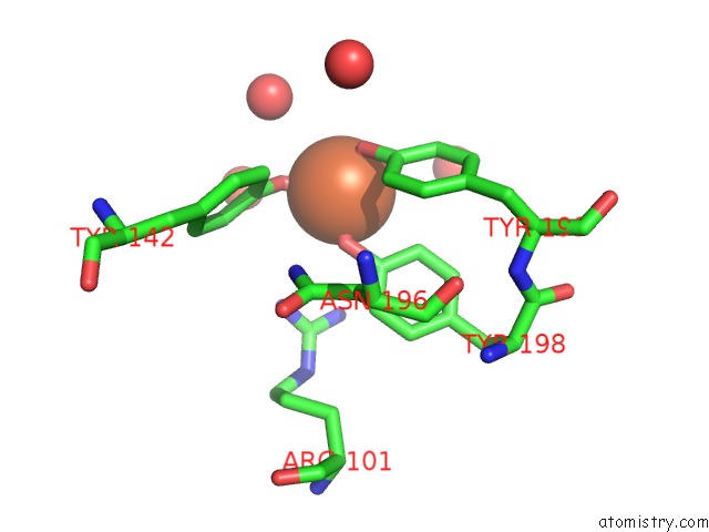
Mono view
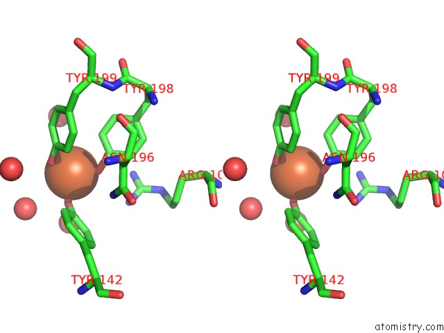
Stereo pair view
Mono view
Stereo pair view
A full contact list of Iron with other atoms in the Fe binding
site number 1 of Crystal Structure of Mannheimia Haemolytica Ferric Iron- Binding Protein A in An Open Conformation within 5.0Å range:
|
Reference:
S.R.Shouldice,
R.J.Skene,
D.R.Dougan,
G.Snell,
D.E.Mcree,
A.B.Schryvers,
L.W.Tari.
Structural Basis For Iron Binding and Release By A Novel Class of Periplasmic Iron-Binding Proteins Found in Gram-Negative Pathogens. J.Bacteriol. V. 186 3903 2004.
ISSN: ISSN 0021-9193
PubMed: 15175304
DOI: 10.1128/JB.186.12.3903-3910.2004
Page generated: Wed Jul 16 20:38:31 2025
ISSN: ISSN 0021-9193
PubMed: 15175304
DOI: 10.1128/JB.186.12.3903-3910.2004
Last articles
Zn in 1KH9Zn in 1KH7
Zn in 1KH5
Zn in 1KH4
Zn in 1KFV
Zn in 1KFS
Zn in 1KEV
Zn in 1KFI
Zn in 1KBP
Zn in 1KEQ