Iron »
PDB 1sdl-1stq »
1sj2 »
Iron in PDB 1sj2: Crystal Structure of Mycobacterium Tuberculosis Catalase-Peroxidase
Enzymatic activity of Crystal Structure of Mycobacterium Tuberculosis Catalase-Peroxidase
All present enzymatic activity of Crystal Structure of Mycobacterium Tuberculosis Catalase-Peroxidase:
1.11.1.6;
1.11.1.6;
Protein crystallography data
The structure of Crystal Structure of Mycobacterium Tuberculosis Catalase-Peroxidase, PDB code: 1sj2
was solved by
T.Bertrand,
N.A.J.Eady,
J.N.Jones,
J.Bodiguel,
Jesmin,
J.M.Nagy,
E.L.Raven,
B.Jamart-Gregoire,
K.A.Brown,
with X-Ray Crystallography technique. A brief refinement statistics is given in the table below:
| Resolution Low / High (Å) | 23.71 / 2.41 |
| Space group | P 42 21 2 |
| Cell size a, b, c (Å), α, β, γ (°) | 150.330, 150.330, 154.281, 90.00, 90.00, 90.00 |
| R / Rfree (%) | 21.1 / 26.8 |
Iron Binding Sites:
The binding sites of Iron atom in the Crystal Structure of Mycobacterium Tuberculosis Catalase-Peroxidase
(pdb code 1sj2). This binding sites where shown within
5.0 Angstroms radius around Iron atom.
In total 2 binding sites of Iron where determined in the Crystal Structure of Mycobacterium Tuberculosis Catalase-Peroxidase, PDB code: 1sj2:
Jump to Iron binding site number: 1; 2;
In total 2 binding sites of Iron where determined in the Crystal Structure of Mycobacterium Tuberculosis Catalase-Peroxidase, PDB code: 1sj2:
Jump to Iron binding site number: 1; 2;
Iron binding site 1 out of 2 in 1sj2
Go back to
Iron binding site 1 out
of 2 in the Crystal Structure of Mycobacterium Tuberculosis Catalase-Peroxidase
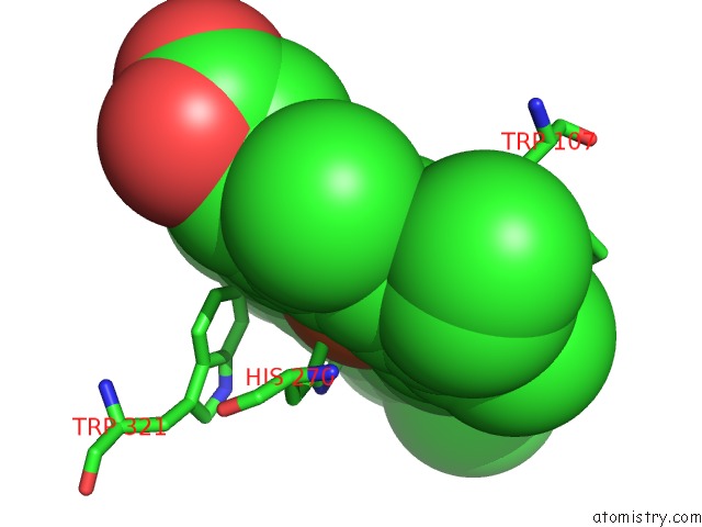
Mono view
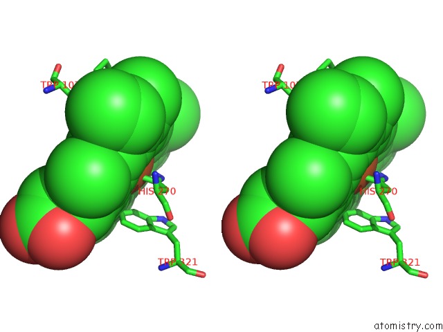
Stereo pair view
Mono view
Stereo pair view
A full contact list of Iron with other atoms in the Fe binding
site number 1 of Crystal Structure of Mycobacterium Tuberculosis Catalase-Peroxidase within 5.0Å range:
|
Iron binding site 2 out of 2 in 1sj2
Go back to
Iron binding site 2 out
of 2 in the Crystal Structure of Mycobacterium Tuberculosis Catalase-Peroxidase
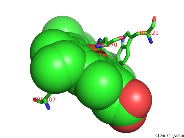
Mono view
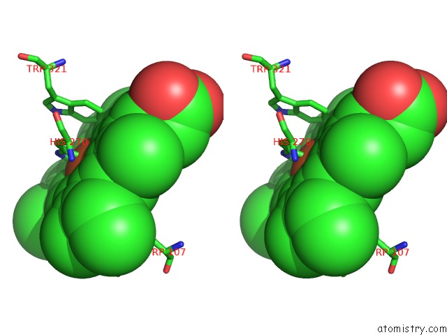
Stereo pair view
Mono view
Stereo pair view
A full contact list of Iron with other atoms in the Fe binding
site number 2 of Crystal Structure of Mycobacterium Tuberculosis Catalase-Peroxidase within 5.0Å range:
|
Reference:
T.Bertrand,
N.A.J.Eady,
J.N.Jones,
Jesmin,
J.M.Nagy,
B.Jamart-Gregoire,
E.L.Raven,
K.A.Brown.
Crystal Structure of Mycobacterium Tuberculosis Catalase-Peroxidase. J.Biol.Chem. V. 279 38991 2004.
ISSN: ISSN 0021-9258
PubMed: 15231843
DOI: 10.1074/JBC.M402382200
Page generated: Wed Jul 16 20:40:27 2025
ISSN: ISSN 0021-9258
PubMed: 15231843
DOI: 10.1074/JBC.M402382200
Last articles
Zn in 1JZQZn in 1JZS
Zn in 1JXP
Zn in 1JYB
Zn in 1JY8
Zn in 1JWH
Zn in 1JWQ
Zn in 1JV0
Zn in 1JWB
Zn in 1JW9